Mouse Model for Colitis ")

- UOM
- FOB US$ 140.00
- Quantity
Overview
Properties
- Product No.DSI515Mu01
- Organism SpeciesMus musculus (Mouse) Same name, Different species.
- ApplicationsDisease Model
Research use only - Downloadn/a
- Category
- Prototype SpeciesHuman
- SourceAcetic acid induced
- Model Animal StrainsBalb/c Mice, healthy, female, 4-6W, body weight 20g-22g
- Modeling GroupingRandomly divided into six group: Control group, Model group, Positive drug group and Test drug group (three doses)
- Modeling Period7d
Sign into your account
Share a new citation as an author
Upload your experimental result
Review

Contact us
Please fill in the blank.
-
 Packages (Simulation)
-
 Packages (Simulation)
-
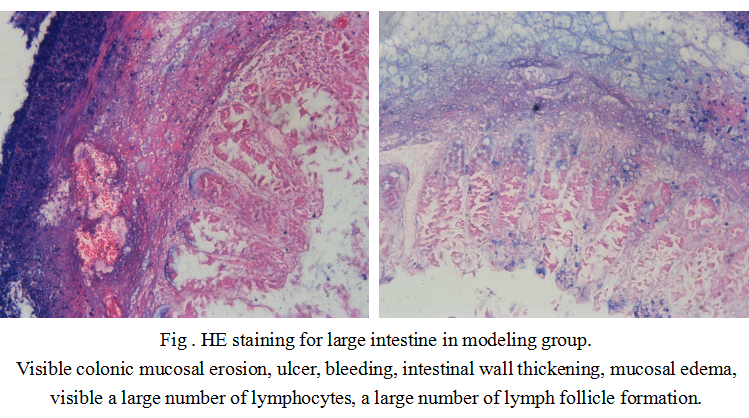 Fig . HE staining for large intestine in modeling group.
-
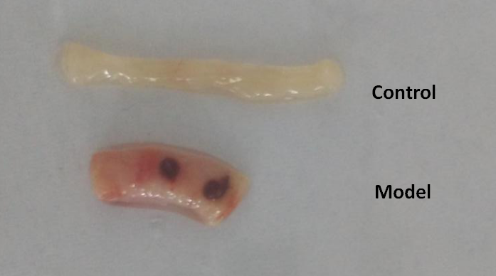 Fig 1. The length of the colon in model group is shorter than that in the control group.
-
 ISO9001: 2008, ISO13485: 2003 Registered
Modeling Method
1.Fasting and free drinking 12 hrs before operation.
2.3% sodium pentobarbital intraperitoneal anesthesia (40 mg/kg) in mice, mice are on the head low tail high position and polyethylene catheter through the anus slowly and insert the 3 cm of colon and the injected 5% acetic acid(0.4 ml), knead tight rats anal and put the mice head down 30s, and then inject saline 1ml, wash 2 times, the control group is injected the normal saline. Mice are allowed to lay flat, natural sober and conventional breeding.
3. 7d after the modeling,take the mice blood, 3000r/min, 4℃, 10min, separate the upper serum, stored at -80℃.
4.Kill mice and seperate the anal end of cecum of the colon and rectum, use the precold saline rinse to wash the colon, dry with filter paper. Observe the changes of colon and measure the length of the colon and record wet weight. Take the most severe lesions of the colon (1 cm), 4% paraformaldehyde, paraffin section, HE staining (4um thick). Observe pathological changes, the rest of the colon are stored at -80℃.
Model evaluation
1.Disease activity index (DAI) :
Observe mice daily diarrhea, bloody stool and body weight changes after modeling, and record the general condition of the rats. After modeling, the body weight of the mice in the model group is decreased significantly, body curled, horripilation, sluggish, loss of appetite, loose stools symptoms.
DAI score: 7 days after modeling, decreased percentage of body weight
No changes for 0 point,
1 ~ 5% for 1 point,
6 ~ 10% for 2 point,
11 ~ 15% for 3 point,
more than 15%, 4 point;
DAI comprehensive score ranged from 0 to 4 points, 0 points on behalf of normal, 4 points on behalf of the maximum degree of inflammation.
Stool viscosity (0 for normal, loose stools for 2 point, diarrhea 4 point);
Stool bleeding (normal 0 and hidden blood positive for 2 point and overt bleeding 4 point.
2.Colon length and colonic mucosal damage index (CMDI):
The length of the colon reflects the indirect indicators of chronic inflammation and injury repair, measuring the length of the anal to the root of the colon is the length of the colon (cm).
Pathological results
Take the most obvious lesions of the colon segment( 1 cm), 4% poly formaldehyde solution fixed, dehydration, paraffin embedded, sliced (thickness of 4 μm), HE staining. HE staining was used to observe the pathological changes in the model group, and the intestinal wall was obviously thickened. Mucosal surfaces have diffuse bleeding spots, erythema, and ulceration. Under the microscope, the mucosa erosion, ulcer, crypt abscess and goblet cells disappeared, and there were neutrophil and lymphocyte infiltration in the lamina. In the normal group, the colonic mucosa of the rats was complete, and the villi arranged neatly, without necrosis or shedding.
Cytokines level
There are a large number of studies have confirmed that free radical levels in the colonic mucosa of ulcerative colitis significantly increased, the lack of antioxidant mechanisms, a large number of free radicals of self-organization produces effect attacks and act as or activation of inflammatory mediators and the inflammatory injury continued to enlarge. The contents of MPO, NO, TNF-α, iNOS, ICAM1 and other cytokines in serum were detected by ELISA kit.
Statistical analysis
SPSS software is used for statistical analysis, measurement data to mean ± standard deviation (x ±s), using t test and single factor analysis of variance for group comparison , P<0.05 indicates there was a significant difference, P<0.01 indicates there are very significant differences.
Giveaways

Increment services
-
 Tissue/Sections Customized Service
-
 Serums Customized Service
-
 Immunohistochemistry (IHC) Experiment Service
-
 Small Animal In Vivo Imaging Experiment Service
-
 Small Animal Micro CT Imaging Experiment Service
-
 Small Animal MRI Imaging Experiment Service
-
 Small Animal Ultrasound Imaging Experiment Service
-
 Transmission Electron Microscopy (TEM) Experiment Service
-
 Scanning Electron Microscope (SEM) Experiment Service
-
 Learning and Memory Behavioral Experiment Service
-
 Anxiety and Depression Behavioral Experiment Service
-
 Drug Addiction Behavioral Experiment Service
-
 Pain Behavioral Experiment Service
-
 Neuropsychiatric Disorder Behavioral Experiment Service
-
 Fatigue Behavioral Experiment Service
-
 Nitric Oxide Assay Kit (A012)
-
 Nitric Oxide Assay Kit (A013-2)
-
 Total Anti-Oxidative Capability Assay Kit(A015-2)
-
 Total Anti-Oxidative Capability Assay Kit (A015-1)
-
 Superoxide Dismutase Assay Kit
-
 Fructose Assay Kit (A085)
-
 Citric Acid Assay Kit (A128 )
-
 Catalase Assay Kit
-
 Malondialdehyde Assay Kit
-
 Glutathione S-Transferase Assay Kit
-
 Microscale Reduced Glutathione assay kit
-
 Glutathione Reductase Activity Coefficient Assay Kit
-
 Angiotensin Converting Enzyme Kit
-
 Glutathione Peroxidase (GSH-PX) Assay Kit
-
 Cloud-Clone Multiplex assay kits