Mouse Model for Tumor Transplantation (TT) ")

Transplanted tumors;TE-1;HepG2;HepG2-luc;SK-MES-1;LL/2-luc-M38
- UOM
- FOB US$ 280.00
- Quantity
Overview
Properties
- Product No.DSI530Mu02
- Organism SpeciesMus musculus (Mouse) Same name, Different species.
- ApplicationsDisease Model for HepG2-luc in situ tumor formation
Research use only - Downloadn/a
- Category
- Prototype SpeciesHuman
- SourceInduced by HepG2-luc in situ tumor formation
- Model Animal StrainsBalb/ -nude Mice(SPF), healthy, male, age:5 weeks, body weight:18g~20g.
- Modeling GroupingRandomly divided into six group: Control group, Model group, Positive drug group and Test drug group(low,medium,high).
- Modeling Period4-6 weeks
Sign into your account
Share a new citation as an author
Upload your experimental result
Review

Contact us
Please fill in the blank.
-
 Packages (Simulation)
-
 Packages (Simulation)
-
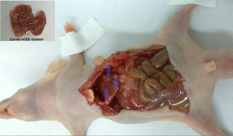 Fig. The liver ( the arrow:tumor on the liver) of nude mice, HepG2-luc cells were injected in situ for 4 weeks
-
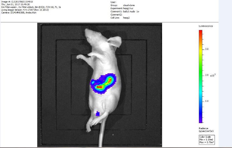 Fig. Fluorescence in vivo imaging after HepG2-luc cells were injected in situ for 1 w
-
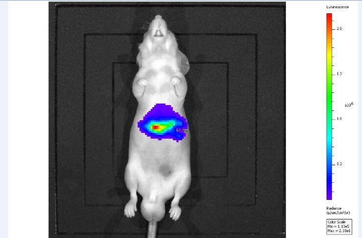 Fig. Fluorescence in vivo imaging after HepG2-luc cells were injected in situ for 1 w
-
 ISO9001: 2008, ISO13485: 2003 Registered
Modeling Method
1. Cell line: HepG2-luc human hepatoma cell line
2. Liver tumor in situ formation model :
2.1 The HepG2-luc cells cultured in logarithmic growth phase were digested and centrifuged, and the resulting approximately 5 x 10e6 cells were dissolved in 0.1 mL DMEM culture medium.
2.2 Use 3% pentobarbital to anesthetize mice, laparotomy to expose the liver, the prepared cells were injected into the liver of nude mice, with 5-0 silk suture wounds, povidone iodine to disinfect wounds, the nude mice were placed in a heating pad, stay awake after normal diet feeding.
3. Fluorescence in vivo imaging:
For in vivo fluorescence imaging detection of tumor in nude mice, mice were anesthetized with 3% pentobarbital sodium used before imaging, by intraperitoneal injection of 3mg luciferin substrate, the substrate after the injection of 20~30 minutes after the in vivo fluorescence imaging detection.
Model evaluation
Pathological results
Cytokines level
Statistical analysis
SPSS software is used for statistical analysis, measurement data to mean ± standard deviation (x ±s), using t test and single factor analysis of variance for group comparison , P<0.05 indicates there was a significant difference, P<0.01 indicates there are very significant differences.
Giveaways

Increment services
-
 Tissue/Sections Customized Service
-
 Serums Customized Service
-
 Immunohistochemistry (IHC) Experiment Service
-
 Small Animal In Vivo Imaging Experiment Service
-
 Small Animal Micro CT Imaging Experiment Service
-
 Small Animal MRI Imaging Experiment Service
-
 Small Animal Ultrasound Imaging Experiment Service
-
 Transmission Electron Microscopy (TEM) Experiment Service
-
 Scanning Electron Microscope (SEM) Experiment Service
-
 Learning and Memory Behavioral Experiment Service
-
 Anxiety and Depression Behavioral Experiment Service
-
 Drug Addiction Behavioral Experiment Service
-
 Pain Behavioral Experiment Service
-
 Neuropsychiatric Disorder Behavioral Experiment Service
-
 Fatigue Behavioral Experiment Service
-
 Nitric Oxide Assay Kit (A012)
-
 Nitric Oxide Assay Kit (A013-2)
-
 Total Anti-Oxidative Capability Assay Kit(A015-2)
-
 Total Anti-Oxidative Capability Assay Kit (A015-1)
-
 Superoxide Dismutase Assay Kit
-
 Fructose Assay Kit (A085)
-
 Citric Acid Assay Kit (A128 )
-
 Catalase Assay Kit
-
 Malondialdehyde Assay Kit
-
 Glutathione S-Transferase Assay Kit
-
 Microscale Reduced Glutathione assay kit
-
 Glutathione Reductase Activity Coefficient Assay Kit
-
 Angiotensin Converting Enzyme Kit
-
 Glutathione Peroxidase (GSH-PX) Assay Kit
-
 Cloud-Clone Multiplex assay kits