Rat Model for Sepsis ")

SIRS; Systemic inflammatory response syndrome
- UOM
- FOB US$ 200.00
- Quantity
Overview
Properties
- Product No.DSI541Ra01
- Organism SpeciesRattus norvegicus (Rat) Same name, Different species.
- ApplicationsDisease Model
Research use only - Downloadn/a
- Category
- Prototype SpeciesHuman
- SourceInduced by cecal ligation and puncture(CLP)
- Model Animal StrainsSD rats (SPF class), healthy, male, body weight 180g~200g
- Modeling GroupingRandomly divided into six group: Control group, Model group, Positive drug group and Test drug group (three doses).
- Modeling Period2d
Sign into your account
Share a new citation as an author
Upload your experimental result
Review

Contact us
Please fill in the blank.
-
 Packages (Simulation)
-
 Packages (Simulation)
-
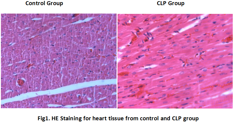 HE Staining for heart tissue from control and CLP group
-
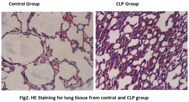 HE Staining for lung tissue from control and CLP group
-
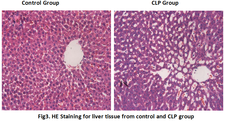 HE Staining for liver tissue from control and CLP group
-
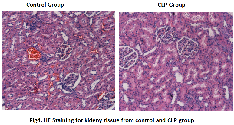 HE Staining for kideny tissue from control and CLP group
-
 ISO9001: 2008, ISO13485: 2003 Registered
Modeling Method
1. Weight the rats, anesthetized by intraperitoneal injection of chloral hydrate (15%) 350 mg/kg, shave the hair on the middle of the head and wipe operation area with tincture of iodine and alcohol.
2. Open abdominal cavity along the linea alba,find out the cecum. Use 5-0 suture to ligate about 1/3 of the cecum. And puncture the ligated cecum 2 times with 21G needle. Gently squeeze small amount of intestinal contents from the puncture hole; put the cecum back into the abdominal cavity, with 5-0 suture, suture the inner layer, 3-0 suture for outer layer. The rats were placed in a heating pad to maintain body temperature at 37±0.5℃.
3. After the rats to wake up free drinking water, normal feeding.
4. After 2 days, the rats were anesthetized, and take 5ml blood from the abdominal aorta, at room temperature for 2hrs, 3000r/min centrifuge 10min at 4℃. Take the heart, liver, kidney, lung and small intestine of the rats, 4% poly formaldehyde fixed, paraffin embedded sections (thickness 4um), HE staining, observe pathological changes, and the rest of the samples stored at -80℃.
Model evaluation
1. General observation
Delayed recovery of postoperative CLP group, after waking, the rats are listlessness, curled up, reduce water consumption, unresponsive to the outside world, respiratory rate, hair fluffy little luster, loose stool, no longer get together.
After 12h, the rats begin to die, along with the extension of time, most of the rats are in low temperature, muscle weakness, eye bloody discharge, stop eating water excretion, watery stool, yellow color, smell. Further development of shortness of breath, no resistance to the passive lying on the back, the discharge was a mucus like. The anatomy of rats showed stench bloody exudate, edema of intestinal adhesions, cecal necrosis black.
Pathological results
Histopathological changes:
(1)Heart: On light microscopy, the control group of myocardial fibers in the structure of the muscle fibers are closely arranged, no edema, congestion and exudation. In model group, the myocardial fibers were arranged loosely, the bands were bubble, the nucleus was swollen, the interstitial edema and congestion.
(2)Liver: In model group, liver has a large number of vacuoles of fatty degeneration, swelling of the liver cells and inflammatory cell infiltration.
(3)Lung: In model group, pulmonary septal thickening, part of the alveolar tissue structure is damaged, inflammatory cell infiltration.
(4)Renal: Renal cortex and interstitial edema with a large number of inflammatory cell infiltration, renal tubular epithelial cells were swelling. Vacuole degeneration, necrosis and shedding. Expansion of renal tubular cyst and tube formation. The state of the cortex and medulla is not clear, the glomerular contraction and capillary micro thrombus formation
(5)Small intestine: intestinal mucosal edema, leukocyte infiltration, hemorrhage and necrosis of epithelial cells in model group.
Cytokines level
The increase of inflammatory cytokines in plasma is one of the typical features of sepsis, IL-1β, TNF-α and IL-6 are the main pro-inflammatory cytokines, and significantly increased in sepsis model.
Statistical analysis
SPSS software is used for statistical analysis, measurement data to mean ± standard deviation (x ±s), using t test and single factor analysis of variance for group comparison , P<0.05 indicates there was a significant difference, P<0.01 indicates there are very significant differences.
Giveaways

Increment services
-
 Tissue/Sections Customized Service
-
 Serums Customized Service
-
 Immunohistochemistry (IHC) Experiment Service
-
 Small Animal In Vivo Imaging Experiment Service
-
 Small Animal Micro CT Imaging Experiment Service
-
 Small Animal MRI Imaging Experiment Service
-
 Small Animal Ultrasound Imaging Experiment Service
-
 Transmission Electron Microscopy (TEM) Experiment Service
-
 Scanning Electron Microscope (SEM) Experiment Service
-
 Learning and Memory Behavioral Experiment Service
-
 Anxiety and Depression Behavioral Experiment Service
-
 Drug Addiction Behavioral Experiment Service
-
 Pain Behavioral Experiment Service
-
 Neuropsychiatric Disorder Behavioral Experiment Service
-
 Fatigue Behavioral Experiment Service
-
 Nitric Oxide Assay Kit (A012)
-
 Nitric Oxide Assay Kit (A013-2)
-
 Total Anti-Oxidative Capability Assay Kit(A015-2)
-
 Total Anti-Oxidative Capability Assay Kit (A015-1)
-
 Superoxide Dismutase Assay Kit
-
 Fructose Assay Kit (A085)
-
 Citric Acid Assay Kit (A128 )
-
 Catalase Assay Kit
-
 Malondialdehyde Assay Kit
-
 Glutathione S-Transferase Assay Kit
-
 Microscale Reduced Glutathione assay kit
-
 Glutathione Reductase Activity Coefficient Assay Kit
-
 Angiotensin Converting Enzyme Kit
-
 Glutathione Peroxidase (GSH-PX) Assay Kit
-
 Cloud-Clone Multiplex assay kits