Rat Model for Traumatic Brain Injury (TBI) ")

Brain Injury
- UOM
- FOB US$ 200.00
- Quantity
Overview
Properties
- Product No.DSI538Ra01
- Organism SpeciesRattus norvegicus (Rat) Same name, Different species.
- ApplicationsDisease Model
Research use only - Downloadn/a
- Category
- Prototype SpeciesHuman
- SourceTraumatic brain strike induced
- Model Animal StrainsSD rats (SPF class), healthy, male, body weight 180g~200g
- Modeling GroupingRandomly divided into six group: Control group, Model group, Positive drug group and Test drug group (three doses).
- Modeling Period7d
Sign into your account
Share a new citation as an author
Upload your experimental result
Review

Contact us
Please fill in the blank.
-
 Packages (Simulation)
-
 Packages (Simulation)
-
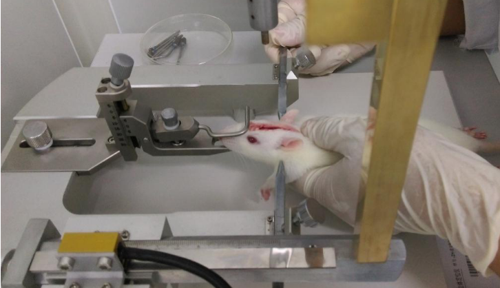 Fig. Operation of Traumatic Brain Injury
-
 ISO9001: 2008, ISO13485: 2003 Registered
Modeling Method
1.Weight the rats, anesthetized, shave the hair on the middle of the head and wipe operation area with tincture of iodine and alcohol.
2. Cut the scalp for 2cm, located at the middle of the head and slightly offset to the right. After blunt separation of soft tissue and bony adventitia, the skull was exposed, 2 mm in front of the herringbone suture and 2 mm beside the midline of the skull. Use skull drilling to open a diameter of 4 mm round bone window, keeping the dura intact. Using the method of free falling impact to brain contusion.
3.A metal object of 40g is fallen from 25cm high vertically.Impacted a cylinder put on the dura mater,impact force is 1000 g/cm. The impact causes right parietal lobe brain contusion and laceration, wound area is 4mm x 4mm, causing severe brain injury, bone window is then blocked with bone wax, then sutured scalp.
4. Free drinking water, normal feeding after the rats to wake up ,
5. Open a window on the right parietal lobe of the corresponding part of the skull, without injury in the control group.
Samples:
1. Blood specimens: after surgery, 72h, 24h and 7d, to take 2ml blood in the inferior vena cava, room temperature static 2h, 3000r centrifuge 10 minutes at 4℃ to extract serum, and stored at -80℃.
2. Brain tissue: take the brain, 4% poly Formaldehyde Solution fixed, after dehydration of sucrose solution, the OCT package to do the frozen sections(10um).
Model evaluation
1.Neurobehavioral score (mNSS score) : a total of 3 days
;Tail lifting experiment; (0-3 points)
The rats were placed on the floor to observe the walking condition. (0-3 points)
Sensory experiment; (0 to 2 points)
Balance beam experiment; (0-6 points)
Reflex loss and abnormal movement tests; (0 to 4 points)
2. Morris water maze
After brain injury, the neurological function of rats is impaired and the cognitive ability is also affected.Morris water maze was used to train rats in each group for 6 days after operation.
3. Brain water content detection:
Independent brain tissue is required to complete the test, and samples cannot be shared with other tests;
Use wet/dry weight ratio.The brain parenchyma (olfactory bulb and posterior cerebellum discarded) was isolated and divided along the midline into two cerebral hemispheres: ipsilateral (right) and contralateral (left). The brain samples were immediately weighed to obtain wet weight (WW) and then dried at 110°C in a drying oven for 24 hours to obtain dry weight (DW). The formula for calculating water content is as follows: Water content (%) = (WW - DW)/WW x 100%.
Pathological results
Cell apoptosis detection(TUNEL)
Traumatic Brain Injury (TBI) produces a large number of apoptotic cells. Apoptosis is occurred in multicellular organisms in a program of cell death process, which a variety of biochemical process will cause changes in cell characteristics, especially on the morphological change, finally leading to cell death. Cell apoptosis can be detected by TUNEL method.
Cytokines level
ELISA: plasma was isolated and the expressions of von Willebrand factor, LBP (Lipopolysaccharide binding protein) and Serpina3n in plasma were detected by ELISA.
Statistical analysis
SPSS software is used for statistical analysis, measurement data to mean ± standard deviation (x ±s), using t test and single factor analysis of variance for group comparison , P<0.05 indicates there was a significant difference, P<0.01 indicates there are very significant differences.
Giveaways

Increment services
-
 Tissue/Sections Customized Service
-
 Serums Customized Service
-
 Immunohistochemistry (IHC) Experiment Service
-
 Small Animal In Vivo Imaging Experiment Service
-
 Small Animal Micro CT Imaging Experiment Service
-
 Small Animal MRI Imaging Experiment Service
-
 Small Animal Ultrasound Imaging Experiment Service
-
 Transmission Electron Microscopy (TEM) Experiment Service
-
 Scanning Electron Microscope (SEM) Experiment Service
-
 Learning and Memory Behavioral Experiment Service
-
 Anxiety and Depression Behavioral Experiment Service
-
 Drug Addiction Behavioral Experiment Service
-
 Pain Behavioral Experiment Service
-
 Neuropsychiatric Disorder Behavioral Experiment Service
-
 Fatigue Behavioral Experiment Service
-
 Nitric Oxide Assay Kit (A012)
-
 Nitric Oxide Assay Kit (A013-2)
-
 Total Anti-Oxidative Capability Assay Kit(A015-2)
-
 Total Anti-Oxidative Capability Assay Kit (A015-1)
-
 Superoxide Dismutase Assay Kit
-
 Fructose Assay Kit (A085)
-
 Citric Acid Assay Kit (A128 )
-
 Catalase Assay Kit
-
 Malondialdehyde Assay Kit
-
 Glutathione S-Transferase Assay Kit
-
 Microscale Reduced Glutathione assay kit
-
 Glutathione Reductase Activity Coefficient Assay Kit
-
 Angiotensin Converting Enzyme Kit
-
 Glutathione Peroxidase (GSH-PX) Assay Kit
-
 Cloud-Clone Multiplex assay kits