Polyclonal Antibody to Complement component 1 Q subcomponent-binding protein, mitochondrial (C1QBP) ")

HABP1; GC1QBP; SF2p32; GC1Q-R; GC1qR; P32; Hyaluronan Binding Protein 1; Splicing Factor SF2-Associated Protein; ASF/SF2-associated p32
Overview
Properties
- Product No.PAA651Hu01
- Organism SpeciesHomo sapiens (Human) Same name, Different species.
- ApplicationsWB,IHC; FCM
If the antibody is used in flow cytometry, please check FCM antibodies.
Research use only - DownloadInstruction Manual
- CategoryImmune molecule
- SourcePolyclonal antibody preparation, Host Rabbit
- Ig Type IgG, Potency n/a
- PurificationAntigen-specific affinity chromatography followed by Protein A affinity chromatography
- LabelNone
- Immunogen RPA651Hu01-Recombinant Complement component 1 Q subcomponent-binding protein, mitochondrial (C1QBP)
- Buffer Formulation0.01M PBS, pH7.4, containing 0.05% Proclin-300, 50% glycerol.
- TraitsLiquid, Concentration 500µg/mL
Sign into your account
Share a new citation as an author
Upload your experimental result
Review

Contact us
Please fill in the blank.
-
 Packages (Simulation)
-
 Packages (Simulation)
-
 DAB staining on IHC-P; Samples: Human Lung cancer Tissue; Primary Ab: 10µg/ml Rabbit Anti-Human HABP1 Antibody Second Ab: 2µg/mL HRP-Linked Caprine Anti-Rabbit IgG Polyclonal Antibody (Catalog: SAA544Rb19)
-
 DAB staining on IHC-P;
Sample: Porcine Colon Tissue
Primary Ab: 10µg/ml Rabbit Anti-Human C1QBP Antibody
Control: Used PBS instead of primary antibody
Second Ab: 2μg/ml HRP-Linked Caprine Anti-Rabbit IgG Polyclonal Antibody
(Catalog: SAA544Rb19) -
 DAB staining on IHC-P;
Samples: Human Prostate cancer Tissue;
Primary Ab: 10µg/ml Rabbit Anti-Human HABP1 Antibody
Second Ab: 2µg/mL HRP-Linked Caprine Anti-Rabbit IgG Polyclonal Antibody
(Catalog: SAA544Rb19) -
 DAB staining on IHC-P;
Sample: Porcine Cerebrum Tissue
Primary Ab: 10µg/ml Rabbit Anti-Human C1QBP Antibody
Control: Used PBS instead of primary antibody
Second Ab: 2μg/ml HRP-Linked Caprine Anti-Rabbit IgG Polyclonal Antibody
(Catalog: SAA544Rb19) -
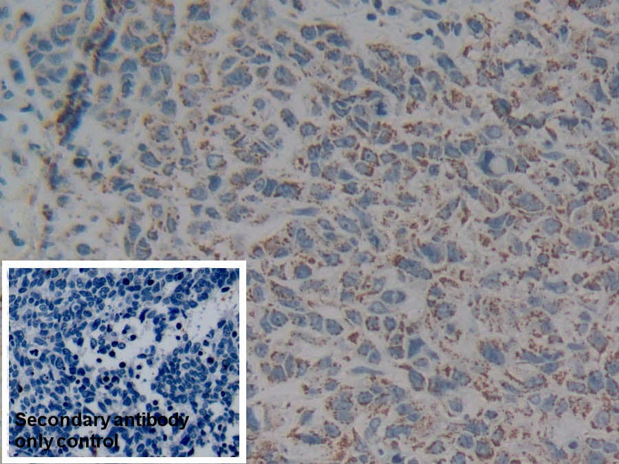
 DAB staining on IHC-P;
Sample: Human Colorectal cancer Tissue;
Primary Ab: 10µg/ml Rabbit Anti-Human HABP1 Antibody
Second Ab: 2µg/mL HRP-Linked Caprine Anti-Rabbit IgG Polyclonal Antibody
(Catalog: SAA544Rb19)
Secondary antibody only control: Used PBS instead of primary antibody, Second Ab: 2µg/mL HRP-Linked Caprine Anti-Rabbit IgG Polyclonal Antibody
(Catalog: SAA544Rb19) -
 DAB staining on IHC-P;
Sample: Mouse Cerebellum Tissue
Primary Ab: 10µg/ml Rabbit Anti-Human C1QBP Antibody
Control: Used PBS instead of primary antibody
Second Ab: 2μg/ml HRP-Linked Caprine Anti-Rabbit IgG Polyclonal Antibody
(Catalog: SAA544Rb19) -
 DAB staining on IHC-P;
Samples: Human Skin cancer Tissue;
Primary Ab: 10µg/ml Rabbit Anti-Human HABP1 Antibody
Second Ab: 2µg/mL HRP-Linked Caprine Anti-Rabbit IgG Polyclonal Antibody
(Catalog: SAA544Rb19) -
 DAB staining on IHC-P;
Samples: Human Pancreatic cancer Tissue;
Primary Ab: 10µg/ml Rabbit Anti-Human HABP1 Antibody
Second Ab: 2µg/mL HRP-Linked Caprine Anti-Rabbit IgG Polyclonal Antibody
(Catalog: SAA544Rb19) -
 DAB staining on IHC-P;
Sample: Mouse Kidney Tissue
Primary Ab: 10µg/ml Rabbit Anti-Human C1QBP Antibody
Control: Used PBS instead of primary antibody
Second Ab: 2μg/ml HRP-Linked Caprine Anti-Rabbit IgG Polyclonal Antibody
(Catalog: SAA544Rb19) -
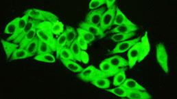
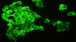
 AF488 staining on IF;
Sample: Hela cell
Primary Ab: 20µg/ml Rabbit Anti-Human C1QBP Antibody
Second Ab: 2μg/ml AF488-Linked Caprine Anti-Rabbit IgG Polyclonal Antibody
(Catalog: SAA544Rb11)
-
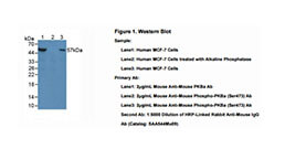
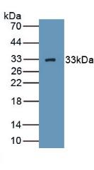 Figure. Western Blot; Sample: Recombinant HABP1, Human.
-
 Western Blot; Sample: Lane1: Mouse Heart lysate; Lane2: Mouse Liver lysate; Lane3: A549 cell lysate; Lane4: Hela cell lysate; Lane5: K562 cell lysate
Primary Ab: 0.1µg/ml Rabbit Anti-Human C1QBP Antibody
Second Ab: 0.2µg/mL HRP-Linked Caprine Anti-Rabbit IgG Polyclonal Antibody
(Catalog: SAA544Rb19) Selected -
 Knockout Varification:
Lane 1: Wild-type Hela cell lysate;
Lane 2: HABP1 knockout Hela cell lysate;
Predicted MW: 31kDa
Observed MW: 31kDa
Primary Ab: 1µg/ml Rabbit Anti-Human HABP1 Antibody
Second Ab: 0.2µg/mL HRP-Linked Caprine Anti-Rabbit IgG Polyclonal Antibody
(Catalog: SAA544Rb19) -
 Human A549 cell was fixed with 2% paraformaldehyde (10 min) , permeabilised with 0.1% BSA-Triton X-100,then stained with 20µg/ml rabbit Anti-human C1QBP Polyclonal Antibody (Catalog PAA651Hu01, red histogram) or Isotype control antibody (Catalog IS067-Rb01, green histogram), followed by 1µg/ml FITC-conjugated Anti-rabbit IgG Secondary Antibody (Catalog SAA544Rb18).
-
 ISO9001: 2008, ISO13485: 2003 Registered
Specifity
The antibody is a rabbit polyclonal antibody raised against C1QBP. It has been selected for its ability to recognize C1QBP in immunohistochemical staining and western blotting.
Usage
Western blotting: 0.01-2μg/mL
Immunohistochemistry: 5-20μg/mL
Flow cytometry:20μg/ml;
Optimal working dilutions must be determined by end user.
Storage
Store at 4°C for frequent use. Stored at -20°C in a manual defrost freezer for two year without detectable loss of activity. Avoid repeated freeze-thaw cycles.
Stability
The thermal stability is described by the loss rate. The loss rate was determined by accelerated thermal degradation test, that is, incubate the protein at 37°C for 48h, and no obvious degradation and precipitation were observed. The loss rate is less than 5% within the expiration date under appropriate storage condition.
Organism Species More: Mus musculus (Mouse)Giveaways
Increment services
-
 Antibody Labeling Customized Service
-
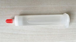 Protein A/G Purification Column
-
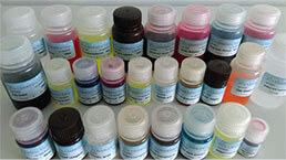 Staining Solution for Cells and Tissue
-
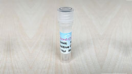 Positive Control for Antibody
-
 Tissue/Sections Customized Service
-
 Phosphorylated Antibody Customized Service
-
 Western Blot (WB) Experiment Service
-
 Immunohistochemistry (IHC) Experiment Service
-
 Immunocytochemistry (ICC) Experiment Service
-
 Flow Cytometry (FCM) Experiment Service
-
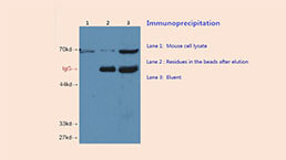 Immunoprecipitation (IP) Experiment Service
-
 Immunofluorescence (IF) Experiment Service
-
 Buffer
-
 DAB Chromogen Kit
-
 SABC Kit
-
 Long-arm Biotin Labeling Kit
-
 Real Time PCR Experimental Service