Recombinant Antibody to Green Fluorescent Protein (GFP) ")

- UOM
- FOB US$ 224.00 US$ 524.00 US$ 748.00 US$ 1,870.00 US$ 7,480.00
- Quantity
Overview
Properties
- Product No.RAD025Ge71
- Organism SpeciesPan-species (General) Same name, Different species.
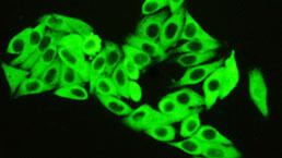
- ApplicationsWB; IF; ICC; IHC; IP; FCM.
If the antibody is used in flow cytometry, please check FCM antibodies.
Research use only - Downloadn/a
- Category
- SourceRecombinant monoclonal antibody preparation, Host CHO
- Ig Isotype IgG, Clone Number n/a
- PurificationProtein A + Protein G affinity chromatography
- LabelBiotin
- Immunogen EPD025Ge51-Eukaryotic Green Fluorescent Protein (GFP)
- Buffer Formulation0.01M PBS, pH7.4, containing 0.05% Proclin-300, 50% glycerol.
- TraitsLiquid, Concentration 1mg/mL
Sign into your account
Share a new citation as an author
Upload your experimental result
Review

Contact us
Please fill in the blank.
-
 Packages (Simulation)
-
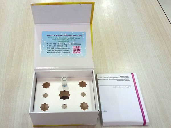 Packages (Simulation)
-
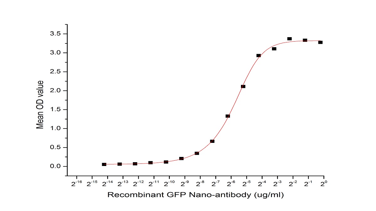 Briefly, biotin labelled GFP Nano-antibody were diluted serially in PBS with 0.01% BSA (pH 7.4). Duplicate samples of 100μl were then transferred to GFP-coated microtiter wells and incubated for 1h at 37℃. Wells were washed 3 times with PBST and incubated for 0.5 h at 37℃ with SA labelled HRP. After incubation with SA labelled HRP, wells were aspirated and washed 5 times. With the addition of substrate solution, wells were incubated 15-25 minutes at 37℃. Finally, add 50µL stop solution to the wells and read at 450/630 nm immediately. The binding activity of GFP and GFP Nano-antibody was shown in Figure 1, and the ED50 for this effect is 0.0187 ug/ml.
-
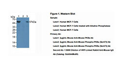
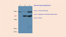
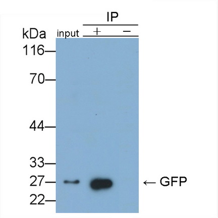 GFP was immunoprecipitated 293F whole cell extract (Expressing GFP) with 2 µg of RAD025Ge71 and 20 µl of Nickel pillar magnetic beads ( ).
Western blot was perfomed on the immunoprecipitate using 0.05µg/ml MAD025Ge21 and 0.2µg/mL SAA544Mu19.
Lane1 (input): 293F whole cell lysate.
Lane2 ( ): RAD025Ge71 in 293F whole cell lysate.
Lane3 (-): No antibody was added to the 293F whole cell lysate.
-
 ISO9001: 2008, ISO13485: 2003 Registered
Specifity
The antibody is a mouse monoclonal antibody raised against GFP. It has been selected for its ability to recognize GFP in immunohistochemical staining and western blotting.
Usage
Western blotting: 0.5-2µg/mL;
Immunohistochemistry: 5-20µg/mL;
Immunocytochemistry: 5-20µg/mL;
Optimal working dilutions must be determined by end user.
Storage
Store at 4°C for frequent use. Stored at -20°C in a manual defrost freezer for two year without detectable loss of activity. Avoid repeated freeze-thaw cycles.
Stability
The thermal stability is described by the loss rate. The loss rate was determined by accelerated thermal degradation test, that is, incubate the protein at 37°C for 48h, and no obvious degradation and precipitation were observed. The loss rate is less than 5% within the expiration date under appropriate storage condition.
Organism Species More: Pan-species (General)Giveaways
Increment services
Citations
- Explore the activation efficiency of different ligand carriers on synNotch-based contact-dependent activation system
- CircPrkcsh, a circular RNA, contributes to the polarization of microglia towards the M1 phenotype induced by spinal cord injury and acts via the JNK/p38 MAPK …34751973
- Japanese Encephalitis Virus NS4A Protein Interacts with PTEN-Induced Kinase 1 (PINK1) and Promotes Mitophagy in Infected CellsPubmed:35604158