Rat Model for Emphysema 

- UOM
- FOB US$ 200.00
- Quantity
Overview
Properties
- Product No.DSI624Ra02
- Organism SpeciesRattus norvegicus (Rat) Same name, Different species.
- ApplicationsDiesase model
Research use only - Downloadn/a
- CategoryRespiratory system
- Prototype SpeciesHuman
- SourceInduced by protease
- Model Animal StrainsSD Rats(SPF), healthy, male and female, body weight 200g~220g.
- Modeling GroupingRandomly divided into six group: Control group, Model group, Positive drug group and Test drug group.
- Modeling Period4-6 weeks
Sign into your account
Share a new citation as an author
Upload your experimental result
Review

Contact us
Please fill in the blank.
Modeling Method
Anesthetized rats with 10% chloral hydrate, after trachea cannula, inject 8% papain (0.5ml/kg BW) with 1ml syringe, maintain the rat upright and revolve it to diffuse drug in lung. Put rats on the heating pad and raise them normally after awake.
The same operation is preformed in control group, but normal saline instead of 8% papain.
Once a day after surgery, rats in model group were given intragastric administration of drugs or normal saline until sampling.
Model evaluation
Each rat is given drug once a week.
A: Control group + normal saline
B: Model control group: Model group + drug
C: Model group + Test drug
Low concentration group;
Middle concentration group;
High concentration group.
D: Positive drug group: Model group + positive drug (Terbutaline sulfate tablets) is given continuously during the duration of rats.
Take several rats from each group at 1W, 2W and 4W after operation, take the left lung for pathological examination.
Serum: Collect whole blood from aorta abdominalis and separate serum, store at -20℃.
Lung homogenate: Take the lung and make tissue homogenate and centrifugation, store at -80℃.
Pathological results
Lung pathology detection:
Take several rats from each group at 1W, 2W and 4W after operation, take left lung after anesthesia and fixed with 4% paraformaldehyde, Then the lung tissue was dehydrated, embedded paraffin section (3μm), HE straining.
Evaluation of lung HE staining can directly reflect the degree of lung injury and judge changes of edema, bleeding, inflammatory cell infiltration, alveolar wall thickness and small airway injury in lung tissue.
Cytokines level
Statistical analysis
SPSS software is used for statistical analysis, measurement data to mean ± standard deviation (x ±s), using t test and single factor analysis of variance for group comparison , P<0.05 indicates there was a significant difference, P<0.01 indicates there are very significant differences.
Giveaways
Increment services
-
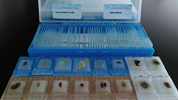 Tissue/Sections Customized Service
-
 Serums Customized Service
-
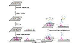 Immunohistochemistry (IHC) Experiment Service
-
 Small Animal In Vivo Imaging Experiment Service
-
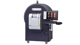 Small Animal Micro CT Imaging Experiment Service
-
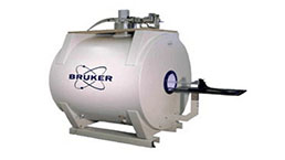 Small Animal MRI Imaging Experiment Service
-
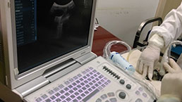 Small Animal Ultrasound Imaging Experiment Service
-
 Transmission Electron Microscopy (TEM) Experiment Service
-
 Scanning Electron Microscope (SEM) Experiment Service
-
 Learning and Memory Behavioral Experiment Service
-
 Anxiety and Depression Behavioral Experiment Service
-
 Drug Addiction Behavioral Experiment Service
-
 Pain Behavioral Experiment Service
-
 Neuropsychiatric Disorder Behavioral Experiment Service
-
 Fatigue Behavioral Experiment Service
-
 Nitric Oxide Assay Kit (A012)
-
 Nitric Oxide Assay Kit (A013-2)
-
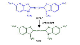 Total Anti-Oxidative Capability Assay Kit(A015-2)
-
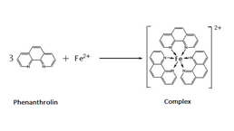 Total Anti-Oxidative Capability Assay Kit (A015-1)
-
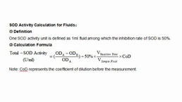 Superoxide Dismutase Assay Kit
-
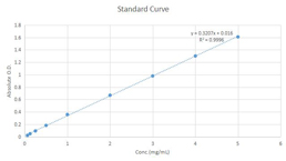 Fructose Assay Kit (A085)
-
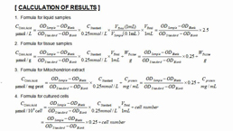 Citric Acid Assay Kit (A128 )
-
 Catalase Assay Kit
-
 Malondialdehyde Assay Kit
-
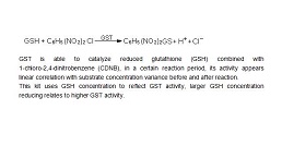 Glutathione S-Transferase Assay Kit
-
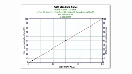 Microscale Reduced Glutathione assay kit
-
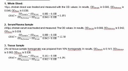 Glutathione Reductase Activity Coefficient Assay Kit
-
 Angiotensin Converting Enzyme Kit
-
 Glutathione Peroxidase (GSH-PX) Assay Kit
-
 Cloud-Clone Multiplex assay kits


