Rat Model for Myocardial Infarction (MI) 

AMI; Acute myocardial infarction; Heart attack
- UOM
- FOB US$
- Quantity
Overview
Properties
- Product No.DSI504Ra01
- Organism SpeciesRattus norvegicus (Rat) Same name, Different species.
- Applicationsn/a
Research use only - Downloadn/a
- CategoryCirculatory system
- Prototype SpeciesHuman
- SourceInduced by monofilament
- Model Animal StrainsWistar Rats(SPF), healthy, male, body weight 300g~400g.
- Modeling Groupingn/a
- Modeling Periodn/a
Sign into your account
Share a new citation as an author
Upload your experimental result
Review

Contact us
Please fill in the blank.
Modeling Method
Use wistar rat with body weight of 300-400g. The diameter of wire is 0.36mm, and the wire can be reused during the experiment. The wire can be shortened to 30cm for easy operation. Shorten The perfusion catheter to 20cm as a cardiac catheter to cause myocardial infarction. Intraperitoneal injection of 3% pentobarbital anesthesia with 50mg/kg. Given a dose of lidocaine(2mg/kg,ip) and heparin(200U/kg,sc) before intubation in order to reduce the formation of ventricular fibrillation and thrombosis. Fix the rabbit supine position, neck median incision. Isolated right common carotid artery, ligated distal. Use A24 subcutaneous injection needle, piercing into the common carotid artery, the success of the piercing standard is arterial blood from the internal needle outflow, when the internal needle removed, the outer casing can slowly into the carotid artery. Exposed carotid artery with wire ligation to prevent blood from flowing out. Cardiac catheter (with j-shaped tip) through the casing under the guidance of fluorescence by the ascending aorta left coronary artery, the wire gently attached to the root of the left coronary artery, the wire counterclockwise rotation into the left coronary artery (LCA). The wire rotates clockwise in the right coronary artery and enters the right coronary artery (RCA). Once the wire into the coronary artery, slowing forward the wire until the animal ECG showed ST segment elevation.
Model evaluation
Pathological results
Cytokines level
Statistical analysis
SPSS software is used for statistical analysis, measurement data to mean ± standard deviation (x ±s), using t test and single factor analysis of variance for group comparison , P<0.05 indicates there was a significant difference, P<0.01 indicates there are very significant differences.
Giveaways
Increment services
-
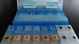 Tissue/Sections Customized Service
-
 Serums Customized Service
-
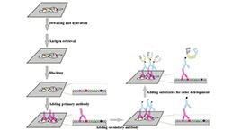 Immunohistochemistry (IHC) Experiment Service
-
 Small Animal In Vivo Imaging Experiment Service
-
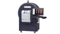 Small Animal Micro CT Imaging Experiment Service
-
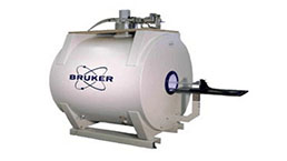 Small Animal MRI Imaging Experiment Service
-
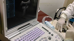 Small Animal Ultrasound Imaging Experiment Service
-
 Transmission Electron Microscopy (TEM) Experiment Service
-
 Scanning Electron Microscope (SEM) Experiment Service
-
 Learning and Memory Behavioral Experiment Service
-
 Anxiety and Depression Behavioral Experiment Service
-
 Drug Addiction Behavioral Experiment Service
-
 Pain Behavioral Experiment Service
-
 Neuropsychiatric Disorder Behavioral Experiment Service
-
 Fatigue Behavioral Experiment Service
-
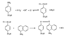 Nitric Oxide Assay Kit (A012)
-
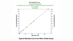 Nitric Oxide Assay Kit (A013-2)
-
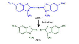 Total Anti-Oxidative Capability Assay Kit(A015-2)
-
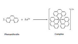 Total Anti-Oxidative Capability Assay Kit (A015-1)
-
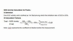 Superoxide Dismutase Assay Kit
-
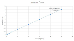 Fructose Assay Kit (A085)
-
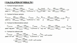 Citric Acid Assay Kit (A128 )
-
 Catalase Assay Kit
-
 Malondialdehyde Assay Kit
-
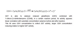 Glutathione S-Transferase Assay Kit
-
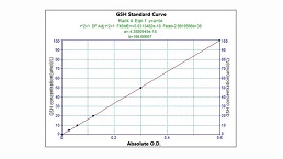 Microscale Reduced Glutathione assay kit
-
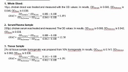 Glutathione Reductase Activity Coefficient Assay Kit
-
 Angiotensin Converting Enzyme Kit
-
 Glutathione Peroxidase (GSH-PX) Assay Kit
-
 Cloud-Clone Multiplex assay kits


