Rat Model for Osteoporosis (OP) ")

- UOM
- FOB US$ 260.00
- Quantity
Overview
Properties
- Product No.DSI534Ra01
- Organism SpeciesRattus norvegicus (Rat) Same name, Different species.
- ApplicationsUsed to study the occurrence process of Osteoporosis, drug intervention and treatment plans
Research use only - Downloadn/a
- Category
- Prototype SpeciesHuman
- SourceInduced by ovariectomy
- Model Animal StrainsWistar Rats(SPF), healthy, male, 6~8 weeks body weight 180g~200g.
- Modeling GroupingRandomly divided into six group: Control group, Model group, Positive drug group and Test drug group.
- Modeling Period6~8 weeks
Sign into your account
Share a new citation as an author
Upload your experimental result
Review

Contact us
Please fill in the blank.
-
 Packages (Simulation)
-
 Packages (Simulation)
-
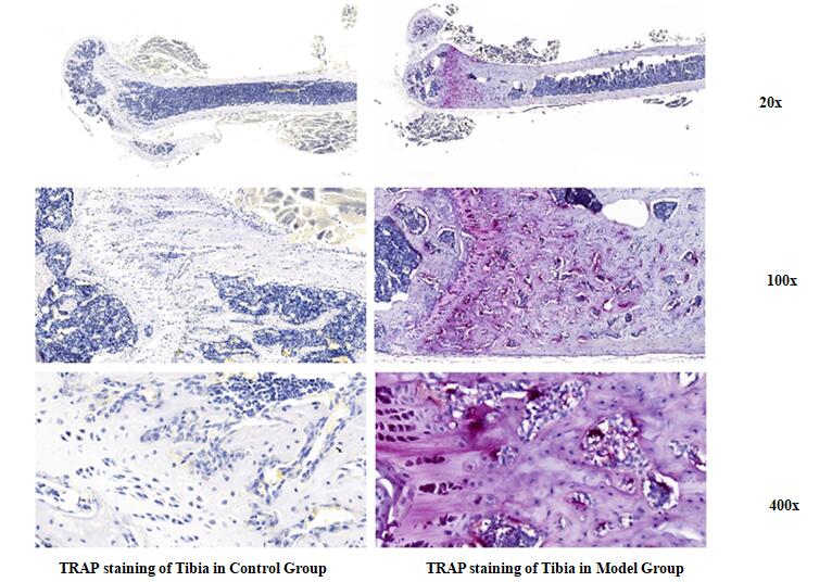 Fig. TRAP staining of tbia in Control and model groupe rats
-
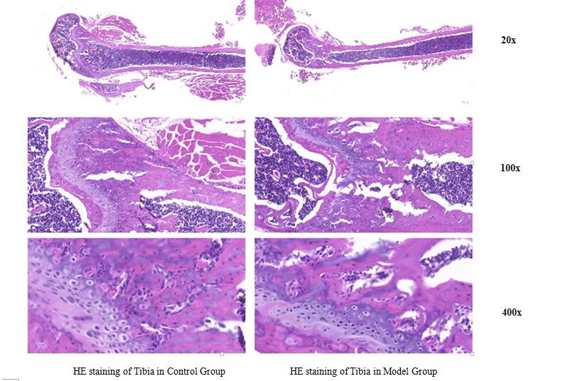 Fig. HE staining of tbia in Control and model groupe rats
-
 ISO9001: 2008, ISO13485: 2003 Registered
Modeling Method
1. Weigh the rats and perform intraperitoneal injection to anesthetize them. After shaving in the middle of the abdomen, wipe the surgical area with iodine and alcohol.
2. Cut open the abdominal cavity along the white line of the abdomen, separate the uterus, and remove the ovaries. After cleaning the wound, sew the skin and base layer in two layers. After the rat wakes up, put it back into a clean cage and return it to the breeding room for feeding. Observe the state and death of the rats and keep records regularly .
3. Rats are injected with 400000 units of penicillin daily to prevent infection three days after surgery,
4. The control group only removed about 1g of fat around the ovaries, while retaining the ovaries. The other treatments are the same.
5. After 8 weeks of surgery, the specimens are executed and collected.
Model evaluation
1. The serum testosterone level of the model group rats is significantly decreased, while the estradiol levels is increased. The biochemical examination results shows that the bone formation indicators of the model group: the serum alkaline phosphatase (ALP) and tartrate resistant acid phosphatase (TRAP) activities of the rats are significantly reduced; The24-hour urine hydroxyproline concentration, creatinine, calcium, and creatinine levels significantly are increased.
Pathological results
Take tibia specimens from each group of rats, fix them for 24-48 hours, and then undergo decalcification. Replace the decalcification solution every 5-7 days; After complete decalcification, gradient ethanol dehydration, xylene transparency, longitudinal paraffin embedding, 5um sectioning, routine HE staining. The tibia of control group rats' trabeculae are dense and interwoven into a network; The number of trabeculae in the model group are significantly decreased, and the trabeculae became thinner and fractured.
Cytokines level
Statistical analysis
SPSS software is used for statistical analysis, measurement data to mean ± standard deviation (x ±s), using t test and single factor analysis of variance for group comparison , P<0.05 indicates there was a significant difference, P<0.01 indicates there are very significant differences.
Giveaways

Increment services
-
 Tissue/Sections Customized Service
-
 Serums Customized Service
-
 Immunohistochemistry (IHC) Experiment Service
-
 Small Animal In Vivo Imaging Experiment Service
-
 Small Animal Micro CT Imaging Experiment Service
-
 Small Animal MRI Imaging Experiment Service
-
 Small Animal Ultrasound Imaging Experiment Service
-
 Transmission Electron Microscopy (TEM) Experiment Service
-
 Scanning Electron Microscope (SEM) Experiment Service
-
 Learning and Memory Behavioral Experiment Service
-
 Anxiety and Depression Behavioral Experiment Service
-
 Drug Addiction Behavioral Experiment Service
-
 Pain Behavioral Experiment Service
-
 Neuropsychiatric Disorder Behavioral Experiment Service
-
 Fatigue Behavioral Experiment Service
-
 Nitric Oxide Assay Kit (A012)
-
 Nitric Oxide Assay Kit (A013-2)
-
 Total Anti-Oxidative Capability Assay Kit(A015-2)
-
 Total Anti-Oxidative Capability Assay Kit (A015-1)
-
 Superoxide Dismutase Assay Kit
-
 Fructose Assay Kit (A085)
-
 Citric Acid Assay Kit (A128 )
-
 Catalase Assay Kit
-
 Malondialdehyde Assay Kit
-
 Glutathione S-Transferase Assay Kit
-
 Microscale Reduced Glutathione assay kit
-
 Glutathione Reductase Activity Coefficient Assay Kit
-
 Angiotensin Converting Enzyme Kit
-
 Glutathione Peroxidase (GSH-PX) Assay Kit
-
 Cloud-Clone Multiplex assay kits