CLIA Kit for Macrophage Inflammatory Protein 3 Alpha (MIP3a) ")

CCL20; CKb4; LARC; MIP3-A; SCYA20; ST38; Small Inducible Cytokine Subfamily A(Cys-Cys)Member 20; Beta-Chemokine Exodus-1; Liver And Activation-Regulated Chemokine
- UOM
- FOB US$ 638.00 US$ 912.00 US$ 4,104.00 US$ 7,752.00 US$ 63,840.00
- Quantity
Overview
Properties
- Product No.SCA095Ra
- Organism SpeciesRattus norvegicus (Rat) Same name, Different species.
- ApplicationsChemiluminescent immunoassay for Antigen Detection.
Research use only - DownloadInstruction Manual
- CategoryCytokineInfection immunity
Sign into your account
Share a new citation as an author
Upload your experimental result
Review

Contact us
Please fill in the blank.
-
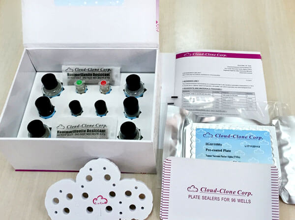
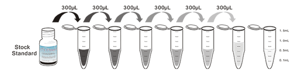
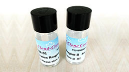
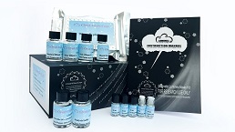
 Packages (Simulation)
-
 Packages (Simulation)
-
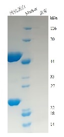
 Results demonstration
-
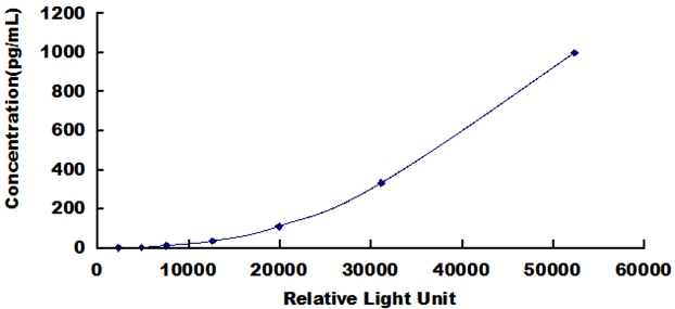 Typical Standard Curve
-
 ISO9001: 2008, ISO13485: 2003 Registered
Recovery
Matrices listed below were spiked with certain level of recombinant Macrophage Inflammatory Protein 3 Alpha (MIP3a) and the recovery rates were calculated by comparing the measured value to the expected amount of Macrophage Inflammatory Protein 3 Alpha (MIP3a) in samples.
| Matrix | Recovery range (%) | Average(%) |
| serum(n=5) | 82-99 | 89 |
| EDTA plasma(n=5) | 80-90 | 85 |
| heparin plasma(n=5) | 92-99 | 96 |
Precision
Intra-assay Precision (Precision within an assay): 3 samples with low, middle and high level Macrophage Inflammatory Protein 3 Alpha (MIP3a) were tested 20 times on one plate, respectively.
Inter-assay Precision (Precision between assays): 3 samples with low, middle and high level Macrophage Inflammatory Protein 3 Alpha (MIP3a) were tested on 3 different plates, 8 replicates in each plate.
CV(%) = SD/meanX100
Intra-Assay: CV<10%
Inter-Assay: CV<12%
Linearity
The linearity of the kit was assayed by testing samples spiked with appropriate concentration of Macrophage Inflammatory Protein 3 Alpha (MIP3a) and their serial dilutions. The results were demonstrated by the percentage of calculated concentration to the expected.
| Sample | 1:2 | 1:4 | 1:8 | 1:16 |
| serum(n=5) | 80-97% | 93-105% | 83-90% | 98-105% |
| EDTA plasma(n=5) | 85-104% | 80-94% | 99-105% | 80-102% |
| heparin plasma(n=5) | 83-101% | 96-104% | 78-104% | 96-105% |
Stability
The stability of kit is determined by the loss rate of activity. The loss rate of this kit is less than 5% within the expiration date under appropriate storage condition.
To minimize extra influence on the performance, operation procedures and lab conditions, especially room temperature, air humidity, incubator temperature should be strictly controlled. It is also strongly suggested that the whole assay is performed by the same operator from the beginning to the end.
Reagents and materials provided
| Reagents | Quantity | Reagents | Quantity |
| Pre-coated, ready to use 96-well strip plate | 1 | Plate sealer for 96 wells | 4 |
| Standard | 2 | Standard Diluent | 1×20mL |
| Detection Reagent A | 1×120µL | Assay Diluent A | 1×12mL |
| Detection Reagent B | 1×120µL | Assay Diluent B | 1×12mL |
| Substrate A | 1×10mL | Substrate B | 1×2mL |
| Wash Buffer (30 × concentrate) | 1×20mL | Instruction manual | 1 |
Assay procedure summary
1. Prepare all reagents, samples and standards;
2. Add 100µL standard or sample to each well. Incubate 1 hours at 37°C;
3. Aspirate and add 100µL prepared Detection Reagent A. Incubate 1 hour at 37°C;
4. Aspirate and wash 3 times;
5. Add 100µL prepared Detection Reagent B. Incubate 30 minutes at 37°C;
6. Aspirate and wash 5 times;
7. Add 100µL Substrate Solution. Incubate 10 minutes at 37°C;
8. Read RLU value immediately.
Test principle
The microplate provided in this kit has been pre-coated with an antibody specific to Macrophage Inflammatory Protein 3 Alpha (MIP3a). Standards or samples are then added to the appropriate microplate wells with a biotin-conjugated antibody specific to Macrophage Inflammatory Protein 3 Alpha (MIP3a). Next, Avidin conjugated to Horseradish Peroxidase (HRP) is added to each microplate well and incubated. Then the mixture of substrate A and B is added to generate glow light emission kinetics. Upon plate development, the intensity of the emitted light is proportional to the Macrophage Inflammatory Protein 3 Alpha (MIP3a) level in the sample or standard.;
Giveaways

Increment services
-
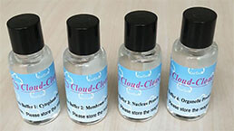
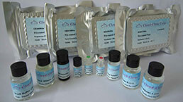 Single-component Reagents of Assay Kit
-
 Lysis Buffer Specific for ELISA / CLIA
-
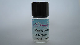 Quality Control of Kit
-
 CLIA Kit Customized Service
-
 Disease Model Customized Service
-
 Serums Customized Service
-
 TGFB1 Activation Reagent
-
 Real Time PCR Experimental Service
-
 Streptavidin
-
 Fast blue Protein Stain solution
-
 Single-component Reagents of FLIA Kit
-
 Streptavidin-Agarose Beads
Citations
- Increased Serum Levels of Macrophage Inflammatory Protein-3α and Cystatin A Predict a Poor Prognosis of Nasopharyngeal CarcinomaPubmed:25396333
- Serum chemokine network correlates with chemotherapy in non-small cell lung cancerPubMed: 25976768
- CCR6+ B lymphocytes responding to tumor cell-derived CCL20 support hepatocellular carcinoma progression via enhancing angiogenesis.pubmed:28560063
- In vitro chemokine (CC motif) receptor 6-dependent non-inflammatory chemotaxis during spermatogenesis10.1186:s40659-018-0161-z
- Indoleamine 2, 3-Dioxygenase (IDO) Regulates Th17/Treg Immunity in Experimental IgA Nephropathy