Multiplex Assay Kit for Activated Protein C (APC) ,etc. by FLIA (Flow Luminescence Immunoassay) ")

(Note: Up to 8-plex in one testing reaction)
- UOM
- FOB US$ 427.00 US$ 443.00 US$ 468.00 US$ 501.00 US$ 534.00 US$ 583.00 US$ 657.00 US$ 821.00
- Quantity
Overview
Properties
- Product No.LMA738Ra
- Organism SpeciesRattus norvegicus (Rat) Same name, Different species.
- ApplicationsFLIA Kit for Antigen Detection.
Research use only - DownloadInstruction Manual
- CategoryEnzyme & KinaseHematology
Sign into your account
Share a new citation as an author
Upload your experimental result
Review

Contact us
Please fill in the blank.
-
 Packages (Simulation)
-
 Packages (Simulation)
-
 Results demonstration
-
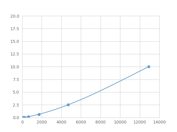 Typical Standard Curve
-
 ISO9001: 2008, ISO13485: 2003 Registered
Recovery
Matrices listed below were spiked with certain level of recombinant Activated Protein C (APC) ,etc. by FLIA (Flow Luminescence Immunoassay) and the recovery rates were calculated by comparing the measured value to the expected amount of Activated Protein C (APC) ,etc. by FLIA (Flow Luminescence Immunoassay) in samples.
| Matrix | Recovery range (%) | Average(%) |
| serum(n=5) | 84-97 | 94 |
| EDTA plasma(n=5) | 94-102 | 98 |
| heparin plasma(n=5) | 86-101 | 96 |
Precision
Intra-assay Precision (Precision within an assay): 3 samples with low, middle and high level Activated Protein C (APC) ,etc. by FLIA (Flow Luminescence Immunoassay) were tested 20 times on one plate, respectively.
Inter-assay Precision (Precision between assays): 3 samples with low, middle and high level Activated Protein C (APC) ,etc. by FLIA (Flow Luminescence Immunoassay) were tested on 3 different plates, 8 replicates in each plate.
CV(%) = SD/meanX100
Intra-Assay: CV<10%
Inter-Assay: CV<12%
Linearity
The linearity of the kit was assayed by testing samples spiked with appropriate concentration of Activated Protein C (APC) ,etc. by FLIA (Flow Luminescence Immunoassay) and their serial dilutions. The results were demonstrated by the percentage of calculated concentration to the expected.
| Sample | 1:2 | 1:4 | 1:8 | 1:16 |
| serum(n=5) | 93-101% | 85-93% | 97-104% | 85-103% |
| EDTA plasma(n=5) | 95-102% | 82-103% | 96-104% | 78-95% |
| heparin plasma(n=5) | 95-103% | 80-99% | 79-93% | 80-96% |
Stability
The stability of kit is determined by the loss rate of activity. The loss rate of this kit is less than 5% within the expiration date under appropriate storage condition.
To minimize extra influence on the performance, operation procedures and lab conditions, especially room temperature, air humidity, incubator temperature should be strictly controlled. It is also strongly suggested that the whole assay is performed by the same operator from the beginning to the end.
Reagents and materials provided
| Reagents | Quantity | Reagents | Quantity |
| 96-well plate | 1 | Plate sealer for 96 wells | 4 |
| Pre-Mixed Standard | 2 | Standard Diluent | 1×20mL |
| Pre-Mixed Magnetic beads (22#:APC) | 1 | Analysis buffer | 1×20mL |
| Pre-Mixed Detection Reagent A | 1×120μL | Assay Diluent A | 1×12mL |
| Detection Reagent B (PE-SA) | 1×120μL | Assay Diluent B | 1×12mL |
| Sheath Fluid | 1×10mL | Wash Buffer (30 × concentrate) | 1×20mL |
| Instruction manual | 1 |
Assay procedure summary
1. Preparation of standards, reagents and samples before the experiment;
2. Add 100μL standard or sample to each well,
add 10μL magnetic beads, and incubate 90min at 37°C on shaker;
3. Remove liquid on magnetic frame, add 100μL prepared Detection Reagent A. Incubate 60min at 37°C on shaker;
4. Wash plate on magnetic frame for three times;
5. Add 100μL prepared Detection Reagent B, and incubate 30 min at 37°C on shaker;
6. Wash plate on magnetic frame for three times;
7. Add 100μL sheath solution, swirl for 2 minutes, read on the machine.

Test principle
Analyte-specific antibodies are pre-coated onto color-coded microparticles. Microparticles, standards, and samples are pipetted into wells and the immobilized antibodies bind the analytes of interest. After washing away any unbound substances, a biotinylated antibody cocktail specific to the analytes of interest is added to each well. Following a wash to remove any unbound biotinylated antibody, Streptavidin-Phycoerythrin conjugate (Streptavidin-PE), which binds to the biotinylated detection antibodies, is added to each well. A final wash removes unbound Streptavidin-PE and the microparticles are resuspended in buffer and read using the Luminex or Bio-Plex analyzer.The MFI developed is proportional to the concentration of analytes of interest in the sample.
Giveaways
Increment services
Citations
- Activated Protein C Attenuates Systemic Lupus Erythematosus and Lupus Nephritis in MRL-Fas(lpr) MiceJimmunol: 3413
- Disseminated intravascular coagulation or acute coagulopathy of trauma shock early after trauma? An observational studyBioMed: cc10553
- Intraovarian Thrombin and Activated Protein C Signaling System Regulates Steroidogenesis during the Periovulatory PeriodEndo: source
- High levels of soluble VEGF receptor 1 early after trauma are associated with shock, sympathoadrenal activation, glycocalyx degradation and inflammation in severely injured patients: a prospective studySjtrem:1757-7241
- Impact of plasma histones in human sepsis and their contribution to cellular injury and inflammationPubmed:25260379
- Monocytes regulate systemic coagulation and inflammation in abdominal sepsisPubmed:25502108
- Protective effects of thrombomodulin on microvascular permeability after subarachnoid hemorrhage in mouse modelPubMed: 25936678
- Activated protein C does not increase in the early phase of trauma with disseminated intravascular coaCavia (Guinea pig )lation: comparison with acute coaCavia (Guinea pig )lopathy of trauma-shockPubmed:26734467
- Brain microvascular endothelial cells exhibit lower activation of the alternative complement pathway than glomerular microvascular endothelial cells.JBC:Source
- A multicenter prospective validation study on disseminated intravascular coagulation in trauma‐induced coagulopathyPubmed: 32480432
- Validation of the Relationship Between Coagulopathy and Localization of Hydroxyethyl Starch on the Vascular Endothelium in a Rat Hemodilution Model34021192