Multiplex Assay Kit for Collagen Type I Alpha 1 (COL1a1) ,etc. by FLIA (Flow Luminescence Immunoassay) ")

COL-1A1; COL1-A1; COL1A-1; OI4; Collagen Alpha-1(I)chain
(Note: Up to 8-plex in one testing reaction)
- UOM
- FOB US$ 393.00 US$ 408.00 US$ 431.00 US$ 461.00 US$ 491.00 US$ 537.00 US$ 605.00 US$ 756.00
- Quantity
Overview
Properties
- Product No.LMA350Hu
- Organism SpeciesHomo sapiens (Human) Same name, Different species.
- ApplicationsFLIA Kit for Antigen Detection.
Research use only - DownloadInstruction Manual
- CategoryMetabolic pathway
Sign into your account
Share a new citation as an author
Upload your experimental result
Review

Contact us
Please fill in the blank.
-
 Packages (Simulation)
-
 Packages (Simulation)
-
 Results demonstration
-
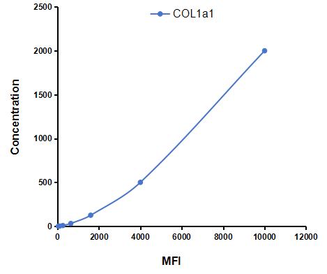 Typical Standard Curve
-
 ISO9001: 2008, ISO13485: 2003 Registered
Recovery
Matrices listed below were spiked with certain level of recombinant Collagen Type I Alpha 1 (COL1a1) ,etc. by FLIA (Flow Luminescence Immunoassay) and the recovery rates were calculated by comparing the measured value to the expected amount of Collagen Type I Alpha 1 (COL1a1) ,etc. by FLIA (Flow Luminescence Immunoassay) in samples.
| Matrix | Recovery range (%) | Average(%) |
| serum(n=5) | 86-101 | 94 |
| EDTA plasma(n=5) | 96-105 | 101 |
| heparin plasma(n=5) | 93-101 | 96 |
| sodium citrate plasma(n=5) | 90-97 | 93 |
Precision
Intra-assay Precision (Precision within an assay): 3 samples with low, middle and high level Collagen Type I Alpha 1 (COL1a1) ,etc. by FLIA (Flow Luminescence Immunoassay) were tested 20 times on one plate, respectively.
Inter-assay Precision (Precision between assays): 3 samples with low, middle and high level Collagen Type I Alpha 1 (COL1a1) ,etc. by FLIA (Flow Luminescence Immunoassay) were tested on 3 different plates, 8 replicates in each plate.
CV(%) = SD/meanX100
Intra-Assay: CV<10%
Inter-Assay: CV<12%
Linearity
The linearity of the kit was assayed by testing samples spiked with appropriate concentration of Collagen Type I Alpha 1 (COL1a1) ,etc. by FLIA (Flow Luminescence Immunoassay) and their serial dilutions. The results were demonstrated by the percentage of calculated concentration to the expected.
| Sample | 1:2 | 1:4 | 1:8 | 1:16 |
| serum(n=5) | 78-94% | 80-95% | 82-90% | 91-98% |
| EDTA plasma(n=5) | 90-97% | 81-104% | 93-105% | 78-98% |
| heparin plasma(n=5) | 92-101% | 93-102% | 95-105% | 85-97% |
| sodium citrate plasma(n=5) | 81-103% | 86-94% | 87-95% | 97-105% |
Stability
The stability of kit is determined by the loss rate of activity. The loss rate of this kit is less than 5% within the expiration date under appropriate storage condition.
To minimize extra influence on the performance, operation procedures and lab conditions, especially room temperature, air humidity, incubator temperature should be strictly controlled. It is also strongly suggested that the whole assay is performed by the same operator from the beginning to the end.
Reagents and materials provided
| Reagents | Quantity | Reagents | Quantity |
| 96-well plate | 1 | Plate sealer for 96 wells | 4 |
| Pre-Mixed Standard | 2 | Standard Diluent | 1×20mL |
| Pre-Mixed Magnetic beads (22#:COL1a1) | 1 | Analysis buffer | 1×20mL |
| Pre-Mixed Detection Reagent A | 1×120μL | Assay Diluent A | 1×12mL |
| Detection Reagent B (PE-SA) | 1×120μL | Assay Diluent B | 1×12mL |
| Sheath Fluid | 1×10mL | Wash Buffer (30 × concentrate) | 1×20mL |
| Instruction manual | 1 |
Assay procedure summary
1. Preparation of standards, reagents and samples before the experiment;
2. Add 100μL standard or sample to each well,
add 10μL magnetic beads, and incubate 90min at 37°C on shaker;
3. Remove liquid on magnetic frame, add 100μL prepared Detection Reagent A. Incubate 60min at 37°C on shaker;
4. Wash plate on magnetic frame for three times;
5. Add 100μL prepared Detection Reagent B, and incubate 30 min at 37°C on shaker;
6. Wash plate on magnetic frame for three times;
7. Add 100μL sheath solution, swirl for 2 minutes, read on the machine.

Test principle
Analyte-specific antibodies are pre-coated onto color-coded microparticles. Microparticles, standards, and samples are pipetted into wells and the immobilized antibodies bind the analytes of interest. After washing away any unbound substances, a biotinylated antibody cocktail specific to the analytes of interest is added to each well. Following a wash to remove any unbound biotinylated antibody, Streptavidin-Phycoerythrin conjugate (Streptavidin-PE), which binds to the biotinylated detection antibodies, is added to each well. A final wash removes unbound Streptavidin-PE and the microparticles are resuspended in buffer and read using the Luminex or Bio-Plex analyzer.The MFI developed is proportional to the concentration of analytes of interest in the sample.
Giveaways
Increment services

Citations
- Simvastatin-loaded porous implant surfaces stimulate preosteoblasts differentiation: an in vitro studyScienceDirect: S1079210410004919
- Osteoblast response to puerarin-loaded porous titanium surfaces: An in vitro studyWiley: source
- Age effects on extracellular matrix production of vocal fold scar fibroblasts in ratsPubmed: 24077847
- Vocal Fold Fibroblast Response to Growth Factor Treatment is Age Dependent: Results From an In Vitro Study Pubmed: 24495429
- Porphyromonas gingivalis LPS inhibits osteoblastic differentiation and promotes pro-inflammatory cytokine production in human periodontal ligament stem cells.Pubmed: 24370188
- Establishing principles of macromolecular crowding for in vitro fibrosis research of the vocal fold lamina propriaPubmed:25545625
- Allogeneic human dermal fibroblasts are viable in peripheral blood mononuclear co-cultureUnivmed:Source
- The vitamin D receptor agonist, calcipotriol, modulates fibrogenic pathways mitigating liver fibrosis in-vivo: An experimental study.pubmed:27477355
- Role of Gut-Derived Endotoxin on Type I Collagen Production in the Rat Pancreas after Chronic Alcohol Exposure pubmed:29121396
- Prevention of Diabetic Nephropathy by Modified Acidic Fibroblast Growth Factorpubmed:28768285
- Effect of TGFβ1, TGFβ3 and keratinocyte conditioned media on functional characteristics of dermal fibroblasts derived from reparative (Balb/c) and …Pubmed:29637306
- LOXL2, a copper-dependent monoamine oxidase, activates lung fibroblasts through the TGF-β/Smad pathwayPubmed: 30320382
- Association between Plasma HMGB-1 and Silicosis: A Case-Control StudyPubmed: 30558126
- Salivary proteins from dysplastic leukoplakia and oral squamous cell carcinoma and their potential for early detectionPubmed: 31706945
- Hesperidin inhibits the epithelial to mesenchymal transition induced by transforming growth factor-β1 in A549 cells through Smad signaling in the cytoplasm
- Platinum nanoparticles supported on functionalized hydroxyapatite: Anti-oxidant properties and bone cells response
- Inhibition of eEF2K synergizes with glutaminase inhibitors or 4EBP1 depletion to suppress growth of triple-negative breast cancer cells33911160
- Antiosteoporotic Nanohydroxyapatite Zoledronate Scaffold Seeded with Bone Marrow Mesenchymal Stromal Cells for Bone Regeneration: A 3D In Vitro ModelPubmed:35682677