Multiplex Assay Kit for Diacylglycerol (DAG) ,etc. by FLIA (Flow Luminescence Immunoassay) ")

DG; Diglyceride
(Note: Up to 8-plex in one testing reaction)
- UOM
- FOB US$ 509.00 US$ 529.00 US$ 558.00 US$ 597.00 US$ 636.00 US$ 695.00 US$ 783.00 US$ 979.00
- Quantity
Overview
Properties
- Product No.LMC038Ge
- Organism SpeciesPan-species (General) Same name, Different species.
- ApplicationsFLIA Kit for Antigen Detection.
Research use only - DownloadInstruction Manual
- CategorySignal transductionEndocrinologyHepatologyGastroenterologyNutrition metabolismHormone metabolism
Sign into your account
Share a new citation as an author
Upload your experimental result
Review

Contact us
Please fill in the blank.
-
 Packages (Simulation)
-
 Packages (Simulation)
-
 Results demonstration
-
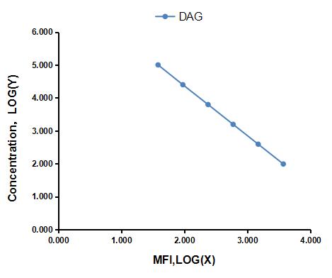 Typical Standard Curve
-
 ISO9001: 2008, ISO13485: 2003 Registered
Recovery
Matrices listed below were spiked with certain level of Diacylglycerol (DAG) ,etc. by FLIA (Flow Luminescence Immunoassay) and the recovery rates were calculated by comparing the measured value to the expected amount of Diacylglycerol (DAG) ,etc. by FLIA (Flow Luminescence Immunoassay) in samples.
| Matrix | Recovery range (%) | Average(%) |
| serum(n=5) | 89-97 | 92 |
| EDTA plasma(n=5) | 87-94 | 90 |
| heparin plasma(n=5) | 84-102 | 98 |
| sodium citrate plasma(n=5) | 84-98 | 91 |
Precision
Intra-assay Precision (Precision within an assay): 3 samples with low, middle and high level Diacylglycerol (DAG) ,etc. by FLIA (Flow Luminescence Immunoassay) were tested 20 times on one plate, respectively.
Inter-assay Precision (Precision between assays): 3 samples with low, middle and high level Diacylglycerol (DAG) ,etc. by FLIA (Flow Luminescence Immunoassay) were tested on 3 different plates, 8 replicates in each plate.
CV(%) = SD/meanX100
Intra-Assay: CV<10%
Inter-Assay: CV<12%
Linearity
The linearity of the kit was assayed by testing samples spiked with appropriate concentration of Diacylglycerol (DAG) ,etc. by FLIA (Flow Luminescence Immunoassay) and their serial dilutions. The results were demonstrated by the percentage of calculated concentration to the expected.
| Sample | 1:2 | 1:4 | 1:8 | 1:16 |
| serum(n=5) | 96-103% | 89-105% | 83-92% | 88-98% |
| EDTA plasma(n=5) | 78-102% | 97-105% | 79-101% | 78-97% |
| heparin plasma(n=5) | 78-102% | 91-98% | 97-104% | 91-101% |
| sodium citrate plasma(n=5) | 92-101% | 88-97% | 85-93% | 88-99% |
Stability
The stability of kit is determined by the loss rate of activity. The loss rate of this kit is less than 5% within the expiration date under appropriate storage condition.
To minimize extra influence on the performance, operation procedures and lab conditions, especially room temperature, air humidity, incubator temperature should be strictly controlled. It is also strongly suggested that the whole assay is performed by the same operator from the beginning to the end.
Reagents and materials provided
| Reagents | Quantity | Reagents | Quantity |
| 96-well plate | 1 | Plate sealer for 96 wells | 4 |
| Pre-Mixed Standard | 2 | Standard Diluent | 1×20mL |
| Pre-Mixed Magnetic beads (22#:DAG) | 1 | Analysis buffer | 1×20mL |
| Pre-Mixed Detection Reagent A | 1×120μL | Assay Diluent A | 1×12mL |
| Detection Reagent B (PE-SA) | 1×120μL | Assay Diluent B | 1×12mL |
| Sheath Fluid | 1×10mL | Wash Buffer (30 × concentrate) | 1×20mL |
| Instruction manual | 1 |
Assay procedure summary
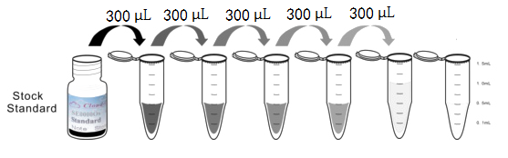
1. Preparation of standards, reagents and samples before the experiment;
2. Add 50μL standard or sample to each well,
add 10μL magnetic beads,and 50μL Detection Reagent A,incubate 60min at 37°C on shaker;
3. Wash plate on magnetic frame for three times;
4. Add 100μL prepared Detection Reagent B, and incubate 30 min at 37°C on shaker;
5. Wash plate on magnetic frame for three times;
6. Add 100μL sheath solution, swirl for 2 minutes, read on the machine.
Test principle
Analyte-specific antibodies are pre-coated onto color-coded microparticles. Microparticles, standards,Labeled antigen and samples are pipetted into wells and the immobilized antibodies bind the analytes of interest.A competitive inhibition reaction is launched between biotin labeled analytes of interest and unlabeled analytes of interest (Standards or samples) with the pre-coated antibody specific to analytes of interest. Following a wash to remove any unbound substances, Streptavidin-Phycoerythrin conjugate (Streptavidin-PE) is added to each well. A final wash removes unbound Streptavidin-PE and the microparticles are resuspended in buffer and read using the Luminex or Bio-Plex analyzer. The MFI developed is reverse proportional to the concentration of analytes of interest in the sample.
Giveaways
Increment services
Citations
- Wogonin ameliorates lipotoxicity-induced apoptosis of cultured vascular smooth muscle cells via interfering with DAG-PKC pathwayNature: 2011120a
- Maternal stress predicts altered biogenesis and the profile of mitochondrial proteins in the frontal cortex and hippocampus of adult offspring ratsPubMed: 26143539
- Aryl Hydrocarbon Receptor Plays Protective Roles against High Fat Diet (HFD)-induced Hepatic Steatosis and the Subsequent Lipotoxicity via Direct Transcriptional ReCavia (Guinea pig )lation of Socs3 Gene ExpressionPubmed:26865635
- Pigment Epithelium-Derived Factor (PEDF) Improves Ischemic Cardiac Functional Reserve Through Decreasing Hypoxic Cardiomyocyte Contractility Through PEDF Receptor (PEDF-R).pubmed:27413044
- PEDF protects cardiomyocytes by promoting FUNDC1‑mediated mitophagy via PEDF-R under hypoxic conditionPubmed:29512692
- Bouchardatine analogue alleviates NAFLD/NASH in high fat fed mice via blunting ATP synthase activityPubmed: 31113010
- Gut Akkermansia muciniphila ameliorates non-alcoholic fatty liver disease by L-aspartate via interaction with liver
- The expression of diacylglycerol kinase isoforms α and ζ correlates with the progression of experimental autoimmune encephalomyelitis in rats34312706