Multiplex Assay Kit for Glucose Transporter 4 (GLUT4) ,etc. by FLIA (Flow Luminescence Immunoassay) ")

SLC2A4; Solute carrier family 2, facilitated glucose transporter member 4; Glucose transporter type 4, insulin-responsive
(Note: Up to 8-plex in one testing reaction)
- UOM
- FOB US$ 449.00 US$ 467.00 US$ 492.00 US$ 527.00 US$ 562.00 US$ 613.00 US$ 691.00 US$ 864.00
- Quantity
Overview
Properties
- Product No.LMC023Mu
- Organism SpeciesMus musculus (Mouse) Same name, Different species.
- ApplicationsFLIA Kit for Antigen Detection.
Research use only - DownloadInstruction Manual
- CategorySignal transductionMetabolic pathwayEndocrinologyCardiovascular biology
Sign into your account
Share a new citation as an author
Upload your experimental result
Review

Contact us
Please fill in the blank.
-
 Packages (Simulation)
-
 Packages (Simulation)
-
 Results demonstration
-
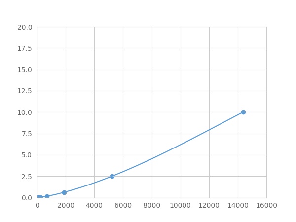 Typical Standard Curve
-
 ISO9001: 2008, ISO13485: 2003 Registered
Recovery
Matrices listed below were spiked with certain level of recombinant Glucose Transporter 4 (GLUT4) ,etc. by FLIA (Flow Luminescence Immunoassay) and the recovery rates were calculated by comparing the measured value to the expected amount of Glucose Transporter 4 (GLUT4) ,etc. by FLIA (Flow Luminescence Immunoassay) in samples.
| Matrix | Recovery range (%) | Average(%) |
| serum(n=5) | 80-95 | 85 |
| EDTA plasma(n=5) | 95-105 | 99 |
| heparin plasma(n=5) | 94-102 | 98 |
Precision
Intra-assay Precision (Precision within an assay): 3 samples with low, middle and high level Glucose Transporter 4 (GLUT4) ,etc. by FLIA (Flow Luminescence Immunoassay) were tested 20 times on one plate, respectively.
Inter-assay Precision (Precision between assays): 3 samples with low, middle and high level Glucose Transporter 4 (GLUT4) ,etc. by FLIA (Flow Luminescence Immunoassay) were tested on 3 different plates, 8 replicates in each plate.
CV(%) = SD/meanX100
Intra-Assay: CV<10%
Inter-Assay: CV<12%
Linearity
The linearity of the kit was assayed by testing samples spiked with appropriate concentration of Glucose Transporter 4 (GLUT4) ,etc. by FLIA (Flow Luminescence Immunoassay) and their serial dilutions. The results were demonstrated by the percentage of calculated concentration to the expected.
| Sample | 1:2 | 1:4 | 1:8 | 1:16 |
| serum(n=5) | 92-104% | 84-92% | 81-96% | 86-94% |
| EDTA plasma(n=5) | 79-96% | 83-90% | 85-101% | 90-97% |
| heparin plasma(n=5) | 86-94% | 96-104% | 85-103% | 98-105% |
Stability
The stability of kit is determined by the loss rate of activity. The loss rate of this kit is less than 5% within the expiration date under appropriate storage condition.
To minimize extra influence on the performance, operation procedures and lab conditions, especially room temperature, air humidity, incubator temperature should be strictly controlled. It is also strongly suggested that the whole assay is performed by the same operator from the beginning to the end.
Reagents and materials provided
| Reagents | Quantity | Reagents | Quantity |
| 96-well plate | 1 | Plate sealer for 96 wells | 4 |
| Pre-Mixed Standard | 2 | Standard Diluent | 1×20mL |
| Pre-Mixed Magnetic beads (22#:GLUT4) | 1 | Analysis buffer | 1×20mL |
| Pre-Mixed Detection Reagent A | 1×120μL | Assay Diluent A | 1×12mL |
| Detection Reagent B (PE-SA) | 1×120μL | Assay Diluent B | 1×12mL |
| Sheath Fluid | 1×10mL | Wash Buffer (30 × concentrate) | 1×20mL |
| Instruction manual | 1 |
Assay procedure summary
1. Preparation of standards, reagents and samples before the experiment;
2. Add 100μL standard or sample to each well,
add 10μL magnetic beads, and incubate 90min at 37°C on shaker;
3. Remove liquid on magnetic frame, add 100μL prepared Detection Reagent A. Incubate 60min at 37°C on shaker;
4. Wash plate on magnetic frame for three times;
5. Add 100μL prepared Detection Reagent B, and incubate 30 min at 37°C on shaker;
6. Wash plate on magnetic frame for three times;
7. Add 100μL sheath solution, swirl for 2 minutes, read on the machine.

Test principle
Analyte-specific antibodies are pre-coated onto color-coded microparticles. Microparticles, standards, and samples are pipetted into wells and the immobilized antibodies bind the analytes of interest. After washing away any unbound substances, a biotinylated antibody cocktail specific to the analytes of interest is added to each well. Following a wash to remove any unbound biotinylated antibody, Streptavidin-Phycoerythrin conjugate (Streptavidin-PE), which binds to the biotinylated detection antibodies, is added to each well. A final wash removes unbound Streptavidin-PE and the microparticles are resuspended in buffer and read using the Luminex or Bio-Plex analyzer.The MFI developed is proportional to the concentration of analytes of interest in the sample.
Giveaways
Increment services

Citations
- Glucose transporter-4 in white blood cells of young and old sled dogs: a model for human biomarker developmentCambridge: Source
- Conditioning causes an increase in glucose transporter-4 levels in mononuclear cells in sled dogsPubmed:25236492
- Maternal stress predicts altered biogenesis and the profile of mitochondrial proteins in the frontal cortex and hippocampus of adult offspring ratsPubMed: 26143539
- Antidiabetic Effect of Galantamine: Novel Effect for a Known Centrally Acting DrugPubMed: 26262991
- Metabolic Features of Heart Failure with Different Etiology ofsp11075
- Hibiscus sabdariffa linn fruits methanolic extract and fractions mediated glucose uptake stimulation and glucose transporter 4 regulationDoi: 10.4103/jpnr.JPNR_14_18
- Effect of Noni Fruit Extract (Morinda citrifolia) on Glucose Intake to Diabetes Mellitus White Rat Muscle Tissue
- The Effects of Sambiloto Leaf Extract (Andrographis peniculata) on Blood Sugar Regulation: an In Vivo Study
- Effect of Bay Leaf Extract (Syzygium polyanthum) on Blood Sugar Regulation via GLUT4 Protein Regulation in White Rat Muscle Tissue Induced Aloxan
- Potential Combination of Tinospora crispa, Andrographis paniculata, Cinamomum burmanii, Syzygium polyanthum and Momordica charantia Extracts Against?¡