


Multiplex Assay Kit for Peroxisome Proliferator Activated Receptor Gamma (PPARg) ,etc. by FLIA (Flow Luminescence Immunoassay) 

PPAR-G; PPARG1; PPARG2; NR1C3; Glitazone Receptor; Nuclear Receptor Subfamily 1 Group C Member 3
(Note: Up to 8-plex in one testing reaction)
- UOM
- FOB US$ 405.00 US$ 420.00 US$ 443.00 US$ 475.00 US$ 506.00 US$ 552.00 US$ 622.00 US$ 778.00
- Quantity
Overview
Properties
- Product No.LMA886Mu
- Organism SpeciesMus musculus (Mouse) Same name, Different species.
- ApplicationsFLIA Kit for Antigen Detection.
Research use only - DownloadInstruction Manual
- CategoryMetabolic pathwayEndocrinologyCardiovascular biologyDevelopmental scienceBone metabolism
Sign into your account
Share a new citation as an author
Upload your experimental result
Review

Contact us
Please fill in the blank.
-
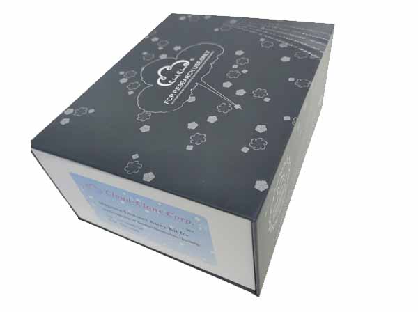 Packages (Simulation)
-
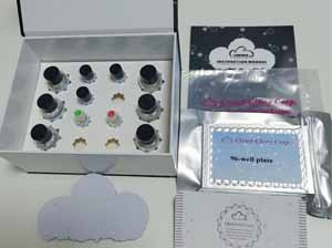 Packages (Simulation)
-
 Results demonstration
-
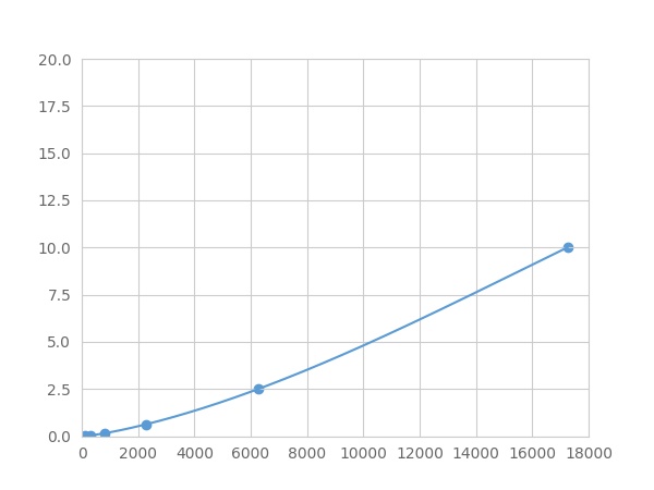 Typical Standard Curve
-
 ISO9001: 2008, ISO13485: 2003 Registered
Recovery
Matrices listed below were spiked with certain level of recombinant Peroxisome Proliferator Activated Receptor Gamma (PPARg) and the recovery rates were calculated by comparing the measured value to the expected amount of Peroxisome Proliferator Activated Receptor Gamma (PPARg) in samples.
| Matrix | Recovery range (%) | Average(%) |
| serum(n=5) | 98-105 | 102 |
| EDTA plasma(n=5) | 93-101 | 96 |
| heparin plasma(n=5) | 79-89 | 86 |
| sodium citrate plasma(n=5) | 90-97 | 93 |
Precision
Intra-assay Precision (Precision within an assay): 3 samples with low, middle and high level Peroxisome Proliferator Activated Receptor Gamma (PPARg) were tested 20 times on one plate, respectively.
Inter-assay Precision (Precision between assays): 3 samples with low, middle and high level Peroxisome Proliferator Activated Receptor Gamma (PPARg) were tested on 3 different plates, 8 replicates in each plate.
CV(%) = SD/meanX100
Intra-Assay: CV<10%
Inter-Assay: CV<12%
Linearity
The linearity of the kit was assayed by testing samples spiked with appropriate concentration of Peroxisome Proliferator Activated Receptor Gamma (PPARg) and their serial dilutions. The results were demonstrated by the percentage of calculated concentration to the expected.
| Sample | 1:2 | 1:4 | 1:8 | 1:16 |
| serum(n=5) | 98-105% | 80-97% | 91-99% | 98-105% |
| EDTA plasma(n=5) | 83-96% | 78-91% | 81-104% | 80-91% |
| heparin plasma(n=5) | 87-101% | 82-103% | 80-97% | 85-99% |
| sodium citrate plasma(n=5) | 95-104% | 98-105% | 90-99% | 84-95% |
Stability
The stability of kit is determined by the loss rate of activity. The loss rate of this kit is less than 5% within the expiration date under appropriate storage condition.
To minimize extra influence on the performance, operation procedures and lab conditions, especially room temperature, air humidity, incubator temperature should be strictly controlled. It is also strongly suggested that the whole assay is performed by the same operator from the beginning to the end.
Reagents and materials provided
| Reagents | Quantity | Reagents | Quantity |
| 96-well plate | 1 | Plate sealer for 96 wells | 4 |
| Pre-Mixed Standard | 2 | Standard Diluent | 1×20mL |
| Pre-Mixed Magnetic beads (22#:PPARg) | 1 | Analysis buffer | 1×20mL |
| Pre-Mixed Detection Reagent A | 1×120μL | Assay Diluent A | 1×12mL |
| Detection Reagent B (PE-SA) | 1×120μL | Assay Diluent B | 1×12mL |
| Sheath Fluid | 1×10mL | Wash Buffer (30 × concentrate) | 1×20mL |
| Instruction manual | 1 |
Assay procedure summary
1. Add 200μL analysis buffer solution to each well of the plate for pre-wetting;
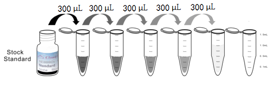
2. Preparation of standards, reagents and samples before the experiment;
3. Add 100μL standard or sample to each well, add 10μL magnetic beads, and incubate 120 minutes protect from light at 25°C on shaker;
4. Remove liquid on magnetic frame, add 100μL prepared Detection Reagent A. Incubate 60 minutes protect from light at 25°C on shaker;
5. Remove liquid on magnetic frame, add 100μL prepared Detection Reagent B, and incubate 30 minutes protect from light at 25°C on shaker;
6. Wash plate once on magnetic frame;
7. Add 100μL sheath fluid, swirl for 10 minutes, read on the machine.
Test principle
Anti-Peroxisome Proliferator Activated Receptor Gamma (PPARg) antibody was coated on the surface of magnetic beads, to make solid phase carrier. Standard or sample were added to the magnetic beads, respectively, in which Peroxisome Proliferator Activated Receptor Gamma (PPARg) was bound to the antibody connected to the solid phase carrier, and then biotinylated anti-Peroxisome Proliferator Activated Receptor Gamma (PPARg) antibodies were added. PE-labeled avidin was added. There is a positive correlation between the concentration of Peroxisome Proliferator Activated Receptor Gamma (PPARg) in the sample and corresponding value of MFI (Median Fluorescence Intensity).
Giveaways
Increment services
Citations
- Elevated levels of PPAR-gamma in the cerebrospinal fluid of patients with multiple sclerosisPubmed: 24021801
- Nonivamide enhances miRNA let‐7d expression and decreases adipogenesis PPARγ expression in 3T3‐L1 cellsPubmed:25704235
- Establishment of a rabbit model to study the influence of advanced glycation end products accumulation on osteoarthritis and the protective effect of pioglitazonePubMed: 26321377
- Evaluation of Protein Kinase Cβ and PPARγ Activity in Diabetic Rats Supplemented with Momordica charantiapmc:PMC4866090
- Evaluation of Protein Kinase Cβ and PPARγ Activity in Diabetic RatsSupplemented with Momordica charantia.pubmed:27190792
- Establishment of a rabbit model to study the influence of advanced glycation end productsaccumulation on osteoarthritis and the protective effect of pioglitazone.pubmed:26321377
- Unlike PPARgamma, neither other PPARs nor PGC-1alpha is elevated in the cerebrospinal fluid of patients with multiple sclerosispubmed:28483651
- Engulfment of Hb‐activated platelets differentiates monocytes into pro‐inflammatory macrophages in PNH patientsPubmed:29677388
- Simpson–Golabi–Behmel syndrome human adipocytes reveal a changing phenotype throughout differentiationPubmed:29574488
- Maternal omega-3 fatty acids and vitamin E improve placental angiogenesis in late-onset but not early-onset preeclampsiaPubmed: 31420792
- Hyperglycemia Changes Expression of Key Adipogenesis Markers (C/EBPα and PPARᵞ) and Morphology of Differentiating Human Visceral AdipocytesPubmed: 31398873
- Indomethacin and juglone inhibit inflammatory molecules to induce apoptosis in colon cancer cellsPubmed: 31916655
- In©\vitro effect of pine bark extract on melanin synthesis, tyrosinase activity, production of endothelin©\1 and PPAR in cultured melanocytes exposed to Ultraviolet?¡33960120
- TRPA1 Agonist Cinnamaldehyde Decreases Adipogenesis in 3T3-L1 Cells More Potently than the Non-agonist Structural Analog Cinnamyl Isobutyrate33403292
- Quantitative real-time measurement of endothelin-1-induced contraction in single non-activated hepatic stellate cells34343209
- Maternal Vitamin D Deficiency Reduces Docosahexaenoic Acid, Placental Growth Factor and Peroxisome Proliferator Activated Receptor Gamma levels in the Pup …34768025
- Melatonin attenuates cisplatin-induced acute kidney injury in mice: Involvement of PPARα and fatty acid oxidationPubmed:35367536