Multiplex Assay Kit for Pigment Epithelium Derived Factor (PEDF) ,etc. by FLIA (Flow Luminescence Immunoassay) ")

SERPINF1; EPC1; PIG35; SDF3; Serpin F1; Cell proliferation-inducing gene 35; Serpin Peptidase Inhibitor,Clade F Member 1(Alpha-2 Antiplasmin); Stromal Cell Derived Factor 3
(Note: Up to 8-plex in one testing reaction)
- UOM
- FOB US$ 427.00 US$ 443.00 US$ 468.00 US$ 501.00 US$ 534.00 US$ 583.00 US$ 657.00 US$ 821.00
- Quantity
Overview
Properties
- Product No.LMB972Mu
- Organism SpeciesMus musculus (Mouse) Same name, Different species.
- ApplicationsFLIA Kit for Antigen Detection.
Research use only - DownloadInstruction Manual
- CategoryCytokineNeuro science
Sign into your account
Share a new citation as an author
Upload your experimental result
Review

Contact us
Please fill in the blank.
-
 Packages (Simulation)
-
 Packages (Simulation)
-
 Results demonstration
-
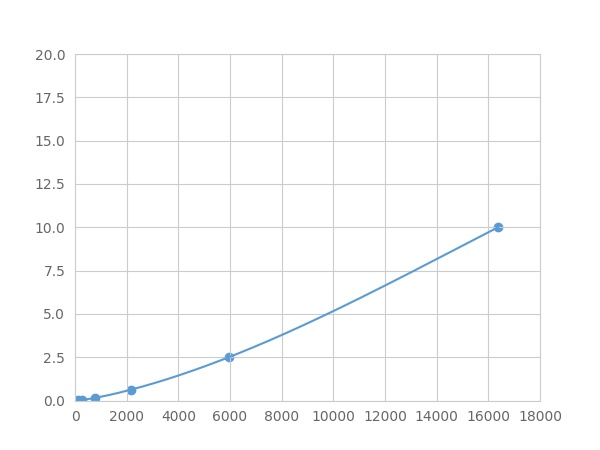 Typical Standard Curve
-
 ISO9001: 2008, ISO13485: 2003 Registered
Recovery
Matrices listed below were spiked with certain level of recombinant Pigment Epithelium Derived Factor (PEDF) ,etc. by FLIA (Flow Luminescence Immunoassay) and the recovery rates were calculated by comparing the measured value to the expected amount of Pigment Epithelium Derived Factor (PEDF) ,etc. by FLIA (Flow Luminescence Immunoassay) in samples.
| Matrix | Recovery range (%) | Average(%) |
| serum(n=5) | 86-95 | 92 |
| EDTA plasma(n=5) | 84-94 | 89 |
| heparin plasma(n=5) | 90-104 | 93 |
Precision
Intra-assay Precision (Precision within an assay): 3 samples with low, middle and high level Pigment Epithelium Derived Factor (PEDF) ,etc. by FLIA (Flow Luminescence Immunoassay) were tested 20 times on one plate, respectively.
Inter-assay Precision (Precision between assays): 3 samples with low, middle and high level Pigment Epithelium Derived Factor (PEDF) ,etc. by FLIA (Flow Luminescence Immunoassay) were tested on 3 different plates, 8 replicates in each plate.
CV(%) = SD/meanX100
Intra-Assay: CV<10%
Inter-Assay: CV<12%
Linearity
The linearity of the kit was assayed by testing samples spiked with appropriate concentration of Pigment Epithelium Derived Factor (PEDF) ,etc. by FLIA (Flow Luminescence Immunoassay) and their serial dilutions. The results were demonstrated by the percentage of calculated concentration to the expected.
| Sample | 1:2 | 1:4 | 1:8 | 1:16 |
| serum(n=5) | 79-91% | 82-101% | 83-101% | 90-104% |
| EDTA plasma(n=5) | 96-103% | 97-104% | 93-105% | 84-101% |
| heparin plasma(n=5) | 80-101% | 80-93% | 95-103% | 97-104% |
Stability
The stability of kit is determined by the loss rate of activity. The loss rate of this kit is less than 5% within the expiration date under appropriate storage condition.
To minimize extra influence on the performance, operation procedures and lab conditions, especially room temperature, air humidity, incubator temperature should be strictly controlled. It is also strongly suggested that the whole assay is performed by the same operator from the beginning to the end.
Reagents and materials provided
| Reagents | Quantity | Reagents | Quantity |
| 96-well plate | 1 | Plate sealer for 96 wells | 4 |
| Pre-Mixed Standard | 2 | Standard Diluent | 1×20mL |
| Pre-Mixed Magnetic beads (22#:PEDF) | 1 | Analysis buffer | 1×20mL |
| Pre-Mixed Detection Reagent A | 1×120μL | Assay Diluent A | 1×12mL |
| Detection Reagent B (PE-SA) | 1×120μL | Assay Diluent B | 1×12mL |
| Sheath Fluid | 1×10mL | Wash Buffer (30 × concentrate) | 1×20mL |
| Instruction manual | 1 |
Assay procedure summary
1. Preparation of standards, reagents and samples before the experiment;
2. Add 100μL standard or sample to each well,
add 10μL magnetic beads, and incubate 90min at 37°C on shaker;
3. Remove liquid on magnetic frame, add 100μL prepared Detection Reagent A. Incubate 60min at 37°C on shaker;
4. Wash plate on magnetic frame for three times;
5. Add 100μL prepared Detection Reagent B, and incubate 30 min at 37°C on shaker;
6. Wash plate on magnetic frame for three times;
7. Add 100μL sheath solution, swirl for 2 minutes, read on the machine.

Test principle
Analyte-specific antibodies are pre-coated onto color-coded microparticles. Microparticles, standards, and samples are pipetted into wells and the immobilized antibodies bind the analytes of interest. After washing away any unbound substances, a biotinylated antibody cocktail specific to the analytes of interest is added to each well. Following a wash to remove any unbound biotinylated antibody, Streptavidin-Phycoerythrin conjugate (Streptavidin-PE), which binds to the biotinylated detection antibodies, is added to each well. A final wash removes unbound Streptavidin-PE and the microparticles are resuspended in buffer and read using the Luminex or Bio-Plex analyzer.The MFI developed is proportional to the concentration of analytes of interest in the sample.
Giveaways
Increment services

Citations
- A Therapeutic Strategy for Choroidal Neovascularization Based on Recruitment of Mesenchymal Stem Cells to the Sites of LesionsNature: 2010144
- Urinary Pigment Epithelium-Derived Factor as a Marker of Diabetic NephropathyKarger: 314326
- Increased expression of pigment epithelium-derived factor in aged mesenchymal stem cells impairs their therapeutic efficacy for attenuating myocardial infarction injuryOxfordJournals: source
- The change of serum level of pigment epithelium-derived factor in coronary heart diseaseBmj: Source
- Metformin Inhibits Expression and Secretion of PEDF in Adipocyte and Hepatocyte via Promoting AMPK PhosphorylationPubmed: 24288442
- The association study of plasma levels of pigment epithelium-derived factor with acute coronary syndrome in the chinese han population.Pubmed: 24192856
- Rosiglitazone Inhibits Expression and Secretion of PEDF in Adipose Tissue and Liver of Male SD Rats Via a PPAR-γ Independent MechanismEndocrine: Source
- Pigment epithelium-derived factor regulates microvascular permeability through adipose triglyceride lipase in sepsisPubMed: 25700221
- Pigment Epithelium-Derived Factor (PEDF) Protects Osteoblastic Cell Line from Glucocorticoid-Induced Apoptosis via PEDF-RPubmed:27187377
- The Ingenious Interactions Between Macrophages and Functionally Plastic Retinal Pigment Epithelium Cellsarticleid:2582822
- VEGF-B promotes recovery of corneal innervations and trophic functions in diabetic micepubmed:28091556
- 色素上皮衍生因子修饰的人脐带间充质干细胞构建方法201707007
- PEDF Reduces the Severity of Herpetic Simplex Keratitis in MicePubmed:30025136
- 高海拔地区新生血管性青光眼不同治疗方式的疗效及对房水中PEDF, VEGF 水平的影响2018/2/201802020.pdf
- Lactation-related changes in tissue expression of PEDF in dairy cowsPubmed:29758402
- Correlation Between Pigment Epithelium-Derived Factor (PEDF) level and Degree of Coronary Angiography and Severity of Coronary Artery Disease in a …Pubmed:29574467
- Role of pigment epithelium-derived factor (PEDF) on arsenic-induced neuronal apoptosisDoi: 10.1016/j.chemosphere.2018.10.100
- Role of Pigment Epithelium-Derived Factor in Arsenic-Induced Vascular Endothelial Dysfunction in a Rat ModelPubmed: 30392020
- Increased levels of serum pigment epithelium-derived factor aggravate proteinuria via induction of podocyte actin rearrangementPubmed: 30536192
- A novel xeno-free culture system for human retinal pigment epithelium cellsPubmed: 31024807
- Pigment Epithelial‐Derived Factor Deficiency Accelerates Atherosclerosis Development via Promoting Endothelial Fatty Acid Uptake in Mice With HyperlipidemiaPubmed: 31711388
- Pigment epithelium-derived factor (PEDF) ameliorates arsenic-induced vascular endothelial dysfunction in rats and toxicity in endothelial EA. hy926 cellsPubmed: 32315827
- The Expression Levels and Cellular Localization of Pigment Epithelium Derived Factor (PEDF) in Mouse Testis: Its Possible Involvement in the Differentiation of Spermatogonial Cells33498962
- Long non-coding RNA Rian promotes the expression of tight junction proteins in endothelial cells by regulating perivascular-resident macrophage-like melanocytes ¡33768511