Multiplex Assay Kit for Pyruvate kinase isozymes M2 (PKM2) ,etc. by FLIA (Flow Luminescence Immunoassay) ")

M2PK; PKM; M2-PK; PKM1; CTHBP; OIP3; PK3; PKM; TCB; THBP1; Pyruvate kinase muscle isozyme; Thyroid hormone-binding protein 1; Cytosolic thyroid hormone-binding protein; Pyruvate Kinase, Muscle
(Note: Up to 8-plex in one testing reaction)
- UOM
- FOB US$ 405.00 US$ 420.00 US$ 443.00 US$ 475.00 US$ 506.00 US$ 552.00 US$ 622.00 US$ 778.00
- Quantity
Overview
Properties
- Product No.LMA588Mu
- Organism SpeciesMus musculus (Mouse) Same name, Different species.
- ApplicationsFLIA Kit for Antigen Detection.
Research use only - DownloadInstruction Manual
- CategoryEnzyme & KinaseTumor immunity
Sign into your account
Share a new citation as an author
Upload your experimental result
Review

Contact us
Please fill in the blank.
-
 Packages (Simulation)
-
 Packages (Simulation)
-
 Results demonstration
-
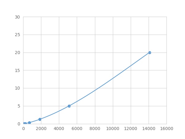 Typical Standard Curve
-
 ISO9001: 2008, ISO13485: 2003 Registered
Recovery
Matrices listed below were spiked with certain level of recombinant Pyruvate kinase isozymes M2 (PKM2) ,etc. by FLIA (Flow Luminescence Immunoassay) and the recovery rates were calculated by comparing the measured value to the expected amount of Pyruvate kinase isozymes M2 (PKM2) ,etc. by FLIA (Flow Luminescence Immunoassay) in samples.
| Matrix | Recovery range (%) | Average(%) |
| serum(n=5) | 84-101 | 95 |
| EDTA plasma(n=5) | 90-97 | 94 |
| heparin plasma(n=5) | 79-90 | 83 |
| sodium citrate plasma(n=5) | 86-96 | 91 |
Precision
Intra-assay Precision (Precision within an assay): 3 samples with low, middle and high level Pyruvate kinase isozymes M2 (PKM2) ,etc. by FLIA (Flow Luminescence Immunoassay) were tested 20 times on one plate, respectively.
Inter-assay Precision (Precision between assays): 3 samples with low, middle and high level Pyruvate kinase isozymes M2 (PKM2) ,etc. by FLIA (Flow Luminescence Immunoassay) were tested on 3 different plates, 8 replicates in each plate.
CV(%) = SD/meanX100
Intra-Assay: CV<10%
Inter-Assay: CV<12%
Linearity
The linearity of the kit was assayed by testing samples spiked with appropriate concentration of Pyruvate kinase isozymes M2 (PKM2) ,etc. by FLIA (Flow Luminescence Immunoassay) and their serial dilutions. The results were demonstrated by the percentage of calculated concentration to the expected.
| Sample | 1:2 | 1:4 | 1:8 | 1:16 |
| serum(n=5) | 94-102% | 98-105% | 79-101% | 97-105% |
| EDTA plasma(n=5) | 78-102% | 80-94% | 93-101% | 85-93% |
| heparin plasma(n=5) | 86-101% | 98-105% | 89-97% | 95-105% |
| sodium citrate plasma(n=5) | 80-104% | 97-105% | 89-103% | 83-96% |
Stability
The stability of kit is determined by the loss rate of activity. The loss rate of this kit is less than 5% within the expiration date under appropriate storage condition.
To minimize extra influence on the performance, operation procedures and lab conditions, especially room temperature, air humidity, incubator temperature should be strictly controlled. It is also strongly suggested that the whole assay is performed by the same operator from the beginning to the end.
Reagents and materials provided
| Reagents | Quantity | Reagents | Quantity |
| 96-well plate | 1 | Plate sealer for 96 wells | 4 |
| Pre-Mixed Standard | 2 | Standard Diluent | 1×20mL |
| Pre-Mixed Magnetic beads (22#:PKM2) | 1 | Analysis buffer | 1×20mL |
| Pre-Mixed Detection Reagent A | 1×120μL | Assay Diluent A | 1×12mL |
| Detection Reagent B (PE-SA) | 1×120μL | Assay Diluent B | 1×12mL |
| Sheath Fluid | 1×10mL | Wash Buffer (30 × concentrate) | 1×20mL |
| Instruction manual | 1 |
Assay procedure summary
1. Preparation of standards, reagents and samples before the experiment;
2. Add 100μL standard or sample to each well,
add 10μL magnetic beads, and incubate 90min at 37°C on shaker;
3. Remove liquid on magnetic frame, add 100μL prepared Detection Reagent A. Incubate 60min at 37°C on shaker;
4. Wash plate on magnetic frame for three times;
5. Add 100μL prepared Detection Reagent B, and incubate 30 min at 37°C on shaker;
6. Wash plate on magnetic frame for three times;
7. Add 100μL sheath solution, swirl for 2 minutes, read on the machine.

Test principle
Analyte-specific antibodies are pre-coated onto color-coded microparticles. Microparticles, standards, and samples are pipetted into wells and the immobilized antibodies bind the analytes of interest. After washing away any unbound substances, a biotinylated antibody cocktail specific to the analytes of interest is added to each well. Following a wash to remove any unbound biotinylated antibody, Streptavidin-Phycoerythrin conjugate (Streptavidin-PE), which binds to the biotinylated detection antibodies, is added to each well. A final wash removes unbound Streptavidin-PE and the microparticles are resuspended in buffer and read using the Luminex or Bio-Plex analyzer.The MFI developed is proportional to the concentration of analytes of interest in the sample.
Giveaways
Increment services

Citations
- Antigen Presentation by Dendritic Cells in Tumors Is Disrupted by Altered Metabolism that Involves Pyruvate Kinase M2 and Its Interaction with SOCS3AACR: 70189
- Abnormal levels of heterogeneous nuclear ribonucleoprotein A2B1 (hnRNPA2B1) in tumour tissue and blood samples from patients diagnosed with lung cancerPubmed:25483567
- M2 isoform of pyruvate kinase (PKM2) is upregulated in Kazakh’s ESCC and promotes proliferation and migration of ESCC cellsPubMed: 26404132
- Identification of a serum biomarker panel for the differential diagnosis of cholangiocarcinoma and primary sclerosing cholangitisPubmed:29707118
- Acteoside improves muscle atrophy and motor function by inducing new myokine secretion in chronic spinal cord injuryPubmed: 30318996
- Secreted Pyruvate Kinase M2 Promotes Lung Cancer Metastasis through Activating the Integrin Beta1/FAK Signaling PathwayPubmed: 32049010
- Chrysin serves as a novel inhibitor of DGKα/FAK interaction to suppress the malignancy of esophageal squamous cell carcinoma (ESCC)
- Bufalin Induced Mitochondrial Dysfunction Promotes Apoptosis of Glioma Cells by Regulating Annexin A2 and DRP1 Proteins
- Bufalin induces mitochondrial dysfunction and promotes apoptosis of glioma cells by regulating Annexin A2 and DRP1 protein expression34376212
- Putative Association between Low Baseline Gene Expression in the Peripheral Blood and Clinical Remission in Rheumatoid Arthritis Patients Treated with Tofacitinib34947916