Multiplex Assay Kit for Tenascin C (TNC) ,etc. by FLIA (Flow Luminescence Immunoassay) ")

TN-C; HXB; TN; GMEM; JI; Myotendinous antigen; Neuronectin; GP 150-225; Cytotactin; Hexabrachion; Glioma-associated-extracellular matrix antigen
(Note: Up to 8-plex in one testing reaction)
- UOM
- FOB US$ 415.00 US$ 431.00 US$ 455.00 US$ 487.00 US$ 519.00 US$ 567.00 US$ 638.00 US$ 798.00
- Quantity
Overview
Properties
- Product No.LMB975Hu
- Organism SpeciesHomo sapiens (Human) Same name, Different species.
- ApplicationsFLIA Kit for Antigen Detection.
Research use only - DownloadInstruction Manual
- CategoryMetabolic pathwayNeuro scienceDevelopmental scienceNutrition metabolismBone metabolism
Sign into your account
Share a new citation as an author
Upload your experimental result
Review

Contact us
Please fill in the blank.
-
 Packages (Simulation)
-
 Packages (Simulation)
-
 Results demonstration
-
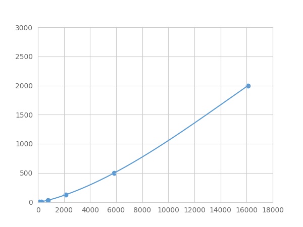 Typical Standard Curve
-
 ISO9001: 2008, ISO13485: 2003 Registered
Recovery
Matrices listed below were spiked with certain level of recombinant Tenascin C (TNC) ,etc. by FLIA (Flow Luminescence Immunoassay) and the recovery rates were calculated by comparing the measured value to the expected amount of Tenascin C (TNC) ,etc. by FLIA (Flow Luminescence Immunoassay) in samples.
| Matrix | Recovery range (%) | Average(%) |
| serum(n=5) | 85-93 | 89 |
| EDTA plasma(n=5) | 81-92 | 84 |
| heparin plasma(n=5) | 93-105 | 97 |
| sodium citrate plasma(n=5) | 80-94 | 85 |
Precision
Intra-assay Precision (Precision within an assay): 3 samples with low, middle and high level Tenascin C (TNC) ,etc. by FLIA (Flow Luminescence Immunoassay) were tested 20 times on one plate, respectively.
Inter-assay Precision (Precision between assays): 3 samples with low, middle and high level Tenascin C (TNC) ,etc. by FLIA (Flow Luminescence Immunoassay) were tested on 3 different plates, 8 replicates in each plate.
CV(%) = SD/meanX100
Intra-Assay: CV<10%
Inter-Assay: CV<12%
Linearity
The linearity of the kit was assayed by testing samples spiked with appropriate concentration of Tenascin C (TNC) ,etc. by FLIA (Flow Luminescence Immunoassay) and their serial dilutions. The results were demonstrated by the percentage of calculated concentration to the expected.
| Sample | 1:2 | 1:4 | 1:8 | 1:16 |
| serum(n=5) | 87-105% | 91-99% | 87-94% | 92-101% |
| EDTA plasma(n=5) | 93-104% | 91-98% | 83-101% | 93-101% |
| heparin plasma(n=5) | 84-103% | 92-101% | 96-104% | 98-105% |
| sodium citrate plasma(n=5) | 78-101% | 81-95% | 78-95% | 88-103% |
Stability
The stability of kit is determined by the loss rate of activity. The loss rate of this kit is less than 5% within the expiration date under appropriate storage condition.
To minimize extra influence on the performance, operation procedures and lab conditions, especially room temperature, air humidity, incubator temperature should be strictly controlled. It is also strongly suggested that the whole assay is performed by the same operator from the beginning to the end.
Reagents and materials provided
| Reagents | Quantity | Reagents | Quantity |
| 96-well plate | 1 | Plate sealer for 96 wells | 4 |
| Pre-Mixed Standard | 2 | Standard Diluent | 1×20mL |
| Pre-Mixed Magnetic beads (22#:TNC) | 1 | Analysis buffer | 1×20mL |
| Pre-Mixed Detection Reagent A | 1×120μL | Assay Diluent A | 1×12mL |
| Detection Reagent B (PE-SA) | 1×120μL | Assay Diluent B | 1×12mL |
| Sheath Fluid | 1×10mL | Wash Buffer (30 × concentrate) | 1×20mL |
| Instruction manual | 1 |
Assay procedure summary
1. Preparation of standards, reagents and samples before the experiment;
2. Add 100μL standard or sample to each well,
add 10μL magnetic beads, and incubate 90min at 37°C on shaker;
3. Remove liquid on magnetic frame, add 100μL prepared Detection Reagent A. Incubate 60min at 37°C on shaker;
4. Wash plate on magnetic frame for three times;
5. Add 100μL prepared Detection Reagent B, and incubate 30 min at 37°C on shaker;
6. Wash plate on magnetic frame for three times;
7. Add 100μL sheath solution, swirl for 2 minutes, read on the machine.

Test principle
Analyte-specific antibodies are pre-coated onto color-coded microparticles. Microparticles, standards, and samples are pipetted into wells and the immobilized antibodies bind the analytes of interest. After washing away any unbound substances, a biotinylated antibody cocktail specific to the analytes of interest is added to each well. Following a wash to remove any unbound biotinylated antibody, Streptavidin-Phycoerythrin conjugate (Streptavidin-PE), which binds to the biotinylated detection antibodies, is added to each well. A final wash removes unbound Streptavidin-PE and the microparticles are resuspended in buffer and read using the Luminex or Bio-Plex analyzer.The MFI developed is proportional to the concentration of analytes of interest in the sample.
Giveaways
Increment services

Citations
- An investigation of tenascin-C levels in rheumatic mitral stenosis and their response to percutaneous mitral balloon valvuloplastyPubmed: 22889719
- Clinical significance of serum tenascin-c levels in epithelial ovarian cancerSpringer: Source
- Clinical significance of serum tenascin-C levels in breast cancerSpringer: Source
- Tenascin-C is increased in atherothrombotic stroke patients and has an anti-inflammatory effect in the human carotid artery.Pubmed:24823872
- Metabolic and cytoprotective effects of in vivo peri-patellar hyaluronic acid injections in cultured tenocytesPubmed:25333747
- Role of Tenascin-C in the differential diagnosis of small round blue cell tumorsIndex.Php: Sdutfd
- A new predictor of mortality in hemodialysis patients; Tenascin-CPubMed: 26390818
- Biomarkers Score for Patients with Mitral Stenosis: A useful conjunction with Wilkins’s Score for Early InterventionHeartresearchopenjournal: Hroj5
- Pankreas Adenokarsinomunda Serum Tenascin-C Düzeyi Potansiyel Bir Biyobelirteç Midir?Cpdf:5C21620161255
- Increased tenascin C and DKK1 in vitiligo: possible role of fibroblasts in acral and non-acral diseasePubmed:29605863
- How to detect and purify tenascin-CPubmed:29310788
- Altered transcriptional regulatory proteins in glioblastoma and YBX1 as a potential regulator of tumor invasionPubmed: 31358880