


Multiplex Assay Kit for Retinoschisin (RS) ,etc. by CBA (Cytometric Bead Array) 

RS1; XLRS1; Retinoschisis X-Linked,Juvenile 1; X-linked juvenile retinoschisis protein
- UOM
- FOB US$ 437.00 US$ 454.00 US$ 479.00 US$ 512.00 US$ 546.00 US$ 596.00 US$ 672.00 US$ 840.00
- Quantity
Overview
Properties
- Product No.CMJ847Hu
- Organism SpeciesHomo sapiens (Human) Same name, Different species.
- ApplicationsFLIA Kit for Antigen Detection.
Research use only - DownloadInstruction Manual
- CategorySignal transduction
Sign into your account
Share a new citation as an author
Upload your experimental result
Review

Contact us
Please fill in the blank.
-
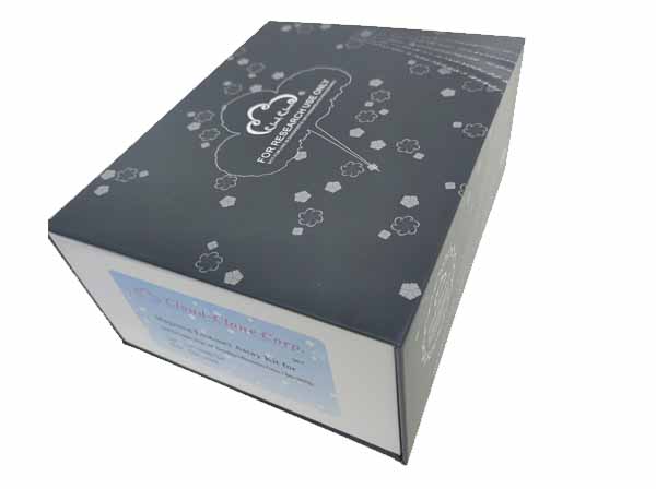 Packages (Simulation)
-
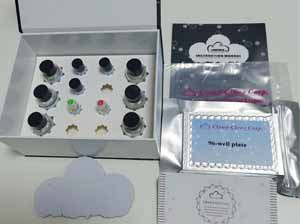 Packages (Simulation)
-
 ISO9001: 2008, ISO13485: 2003 Registered
Recovery
Matrices listed below were spiked with certain level of recombinant Retinoschisin (RS) ,etc. by CBA (Cytometric Bead Array) and the recovery rates were calculated by comparing the measured value to the expected amount of Retinoschisin (RS) ,etc. by CBA (Cytometric Bead Array) in samples.
| Matrix | Recovery range (%) | Average(%) |
| serum(n=5) | 79-103 | 92 |
| EDTA plasma(n=5) | 82-90 | 87 |
| heparin plasma(n=5) | 95-102 | 99 |
Precision
Intra-assay Precision (Precision within an assay): 3 samples with low, middle and high level Retinoschisin (RS) ,etc. by CBA (Cytometric Bead Array) were tested 20 times on one plate, respectively.
Inter-assay Precision (Precision between assays): 3 samples with low, middle and high level Retinoschisin (RS) ,etc. by CBA (Cytometric Bead Array) were tested on 3 different plates, 8 replicates in each plate.
CV(%) = SD/meanX100
Intra-Assay: CV<10%
Inter-Assay: CV<12%
Linearity
The linearity of the kit was assayed by testing samples spiked with appropriate concentration of Retinoschisin (RS) ,etc. by CBA (Cytometric Bead Array) and their serial dilutions. The results were demonstrated by the percentage of calculated concentration to the expected.
| Sample | 1:2 | 1:4 | 1:8 | 1:16 |
| serum(n=5) | 79-90% | 95-103% | 91-105% | 86-98% |
| EDTA plasma(n=5) | 84-96% | 93-101% | 80-93% | 96-104% |
| heparin plasma(n=5) | 88-105% | 92-105% | 86-102% | 94-101% |
Stability
The stability of kit is determined by the loss rate of activity. The loss rate of this kit is less than 5% within the expiration date under appropriate storage condition.
To minimize extra influence on the performance, operation procedures and lab conditions, especially room temperature, air humidity, incubator temperature should be strictly controlled. It is also strongly suggested that the whole assay is performed by the same operator from the beginning to the end.
Reagents and materials provided
| Reagents | Quantity | Reagents | Quantity |
Assay procedure summary
1. Add 200μL analysis buffer solution to each well of the plate for pre-wetting;
2. Preparation of standards, reagents and samples before the experiment;
3. Add 100μL standard or sample to each well, add 10μL magnetic beads, and incubate 120 minutes protect from light at 25°C on shaker;
4. Remove liquid on magnetic frame, add 100μL prepared Detection Reagent A. Incubate 60 minutes protect from light at 25°C on shaker;
5. Remove liquid on magnetic frame, add 100μL prepared Detection Reagent B, and incubate 30 minutes protect from light at 25°C on shaker;
6. Wash plate once on magnetic frame;
7. Add 100μL sheath fluid, swirl for 10 minutes, read on the machine.
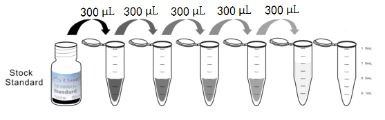
Test principle
Anti-Retinoschisin (RS) ,etc. by CBA (Cytometric Bead Array) antibody was coated on the surface of magnetic beads, to make solid phase carrier. Standard or sample were added to the magnetic beads, respectively, in which Retinoschisin (RS) ,etc. by CBA (Cytometric Bead Array) was bound to the antibody connected to the solid phase carrier, and then biotinylated anti-Retinoschisin (RS) ,etc. by CBA (Cytometric Bead Array) antibodies were added. PE-labeled avidin was added. There is a positive correlation between the concentration of Retinoschisin (RS) ,etc. by CBA (Cytometric Bead Array) in the sample and corresponding value of MFI (Median Fluorescence Intensity). Flow Cytometer was used to determine and calculate the sample concentration.
Giveaways
Increment services
Citations
- Dextran and protamine-based solid lipid nanoparticles as potential vectors for the treatment of X-linked juvenile retinoschisis.PubMed: 22295905
- Influence of Iron Oxide Nanoparticles on Innate and Genetically Modified Secretion Profiles of Mesenchymal Stem CellsScholarsportal: Source
- A novel gene therapy vector based on hyaluronic acid and solid lipid nanoparticles for ocular diseasesPubmed:24576595
- Structural recovery of the retina in a retinoschisin-deficient mouse after gene replacement therapy by solid lipid nanoparticlesPubmed:26986855
- Cell based therapeutics for retinal degenerationscIRcle:1.0303136
- An ex vivo gene therapy approach in X-linked retinoschisispubmed:27390514