Active Colony Stimulating Factor 3, Granulocyte (GCSF) ")

CSF3; G-CSF; Granulocyte Colony Stimulating Factor; Pluripoietin; Filgrastim; Lenograstim
- UOM
- FOB US$ 292.00 US$ 730.00 US$ 1,460.00 US$ 4,380.00 US$ 10,950.00
- Quantity
Overview
Properties
- Product No.APA042Po01
- Organism SpeciesSus scrofa; Porcine (Pig) Same name, Different species.
- ApplicationsCell culture; Activity Assays.
Research use only - DownloadInstruction Manual
- CategoryCytokineHematology
- Buffer FormulationPBS, pH7.4, containing 0.01% SKL, 5% Trehalose.
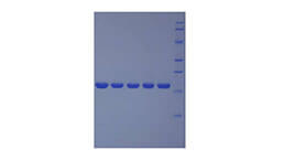
- Traits Freeze-dried powder, Purity > 95%
- Isoelectric Point6.3
Sign into your account
Share a new citation as an author
Upload your experimental result
Review

Contact us
Please fill in the blank.
-
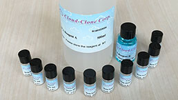
 Packages (Simulation)
-
 Packages (Simulation)
-
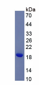 Figure. SDS-PAGE
-
 ISO9001: 2008, ISO13485: 2003 Registered
Activity test
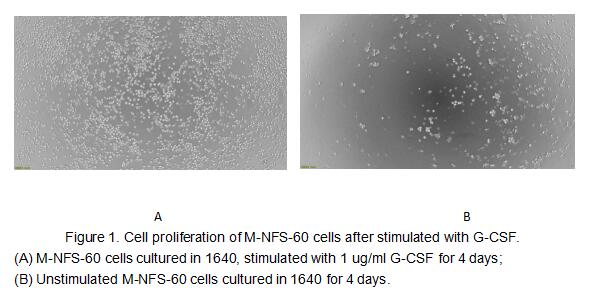
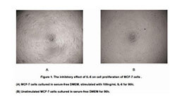
G-CSF is a pleiotropic cytokine best known for its specific effects on the proliferation, differentiation, and activation of hematopoietic cells of the neutrophilic granulocyte lineage. It is produced mainly by monocytes and macrophages upon activation by endotoxin, TNF-alpha and IFN-gamma. In addition, various carcinoma cell lines and myeloblastic leukemia cells can express G-CSF constitutively. In vitro, G-CSF stimulates growth, differentiation and functions of cells from the neutrophil lineage. Consistent with its in vitro functions, G-CSF has been found to play important roles in defense against infection, in inflammation and repair, and in the maintenance of steady state hematopoiesis. The activity of G-CSF is usually measured by a cell proliferation assay using M-NFS 60 mouse myelogenous leukemia lymphoblast cells. M-NFS 60 cells were seeded into triplicate wells of 96-well plates at a density of 8,000 cells/well with 2% serum standard 1640 which contains various concentrations of recombinant pig G-CSF. After incubated for 4 days, cells were observed by inverted microscope and cell proliferation was measured by Cell Counting Kit-8 (CCK-8). Briefly, 10 µl of CCK-8 solution was added to each well of the plate, then the absorbance at 450 nm was measured using a microplate reader after incubating the plate for 2-4 hours at 37 ℃. Proliferation of M-NFS-60 cells after incubation with G-CSF for 4 days observed by inverted microscope was shown in Figure 1. Cell viability was assessed by CCK-8 (Cell Counting Kit-8 ) assay after incubation with recombinant pig G-CSF for 4 days. The result was shown in Figure 2. It was obvious that G-CSF significantly increased cell viability of M-NFS-60 cells. The EC50 is 1 ug/ml
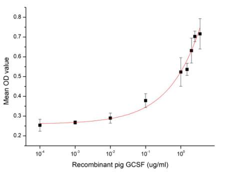
Figure 2. Cell proliferation of M-NFS-60 cells after stimulated with G-CSF.
Usage
Reconstitute in 10mM PBS (pH7.4) to a concentration of 0.1-1.0 mg/mL. Do not vortex.
Storage
Avoid repeated freeze/thaw cycles. Store at 2-8°C for one month. Aliquot and store at -80°C for 12 months.
Stability
The thermal stability is described by the loss rate. The loss rate was determined by accelerated thermal degradation test, that is, incubate the protein at 37°C for 48h, and no obvious degradation and precipitation were observed. The loss rate is less than 5% within the expiration date under appropriate storage condition.
Increment services
-
 BCA Protein Quantification Kit
-
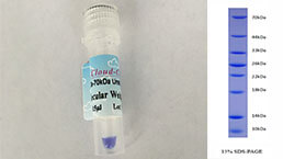 Molecular Mass Marker for Protein
-
 Monoclonal Antibody Customized Service
-
 Polyclonal Antibody Customized Service
-
 Protein Activity Test Experiment Service
-
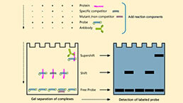 Electrophoretic Mobility Shift Assay (EMSA) Experiment Service
-
 Buffer
-
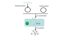
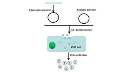 Lentivirus Packaging Experiment Service
-
 Adenovirus Packaging Experiment Service
-
 Real Time PCR Experimental Service
-
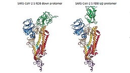 Spike RBD Protein (S-RBD)
-
 Protein G
-
 Protein A
Citations
- The Transcriptional Foundations of Sp110-mediated Macrophage (RAW264. 7) Resistance to Mycobacterium tuberculosis H37RaPubmed:26912204
- Low-density neutrophils in severe fever with thrombocytopenia syndrome (SFTS) display decreased function to phagocytose SFTS virus and enhanced capacity to synthesize cytokinesISSN:1936-2625/IJCEP0045334
- Ginsenoside Rg3 improves cyclophosphamide-induced immunocompetence in Balb/c micePubmed: 30974284
- Oridonin alleviates kanamycin-related hearing loss by inhibiting NLRP3/caspase-1/gasdermin D-induced inflammasome activation and hair cell pyroptosisPubmed:35749835