Active Fibroblast Growth Factor 1, Acidic (FGF1) ")

ECGF; AFGF; ECGFA; ECGFB; FGF-Alpha; FGFA; HBGF1; ECGFB; HBGF1; Acidic Fibroblast Growth Factor; heparin-binding growth factor 1; Endothelial Cell Growth Factor, Beta
Overview
Properties
- Product No.APA032Mu01
- Organism SpeciesMus musculus (Mouse) Same name, Different species.
- ApplicationsCell culture; Activity Assays.
Research use only - DownloadInstruction Manual
- CategoryCytokineTumor immunityInfection immunity
- Buffer Formulation20mM Tris, 150mM NaCl, pH8.0, containing 1mM EDTA, 1mM DTT, 0.01% SKL, 5% Trehalose and Proclin300.
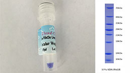
- Traits Freeze-dried powder, Purity > 97%
- Isoelectric Point7.2
Sign into your account
Share a new citation as an author
Upload your experimental result
Review

Contact us
Please fill in the blank.
-
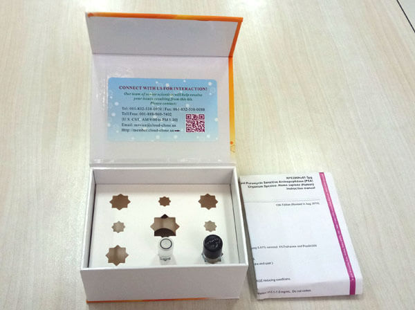
 Packages (Simulation)
-
 Packages (Simulation)
-
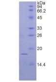 SDS-PAGE
-
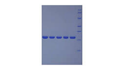
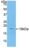 Figure. Western Blot; Sample: Recombinant FGF1, Mouse.
-
 ISO9001: 2008, ISO13485: 2003 Registered
Activity test
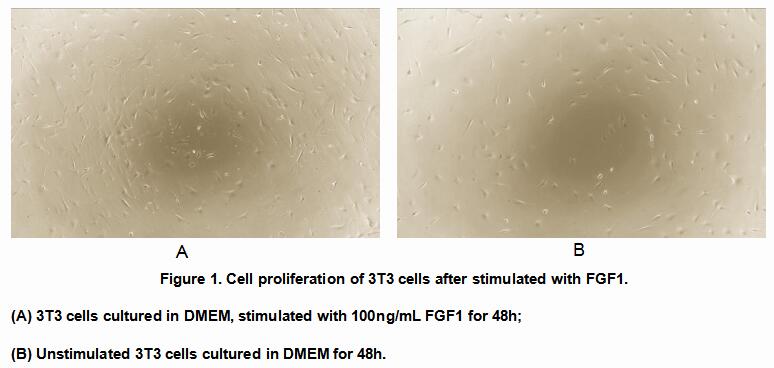
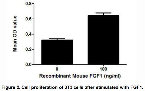
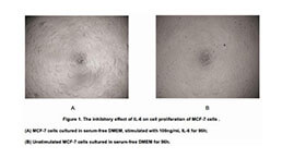
(FGF1) Fibroblast growth factor 1 belongs to the fibroblast growth factor (FGF) family. FGF1 plays an important role in the regulation of cell survival, cell division, angiogenesis, cell differentiation and cell migration. FGF1 is thought to stimulate the proliferation of 3T3 fibroblasts. Thus, a cell proliferation assay was conducted to detect the bioactivity of recombinant mouse FGF1 using 3T3 fibroblasts. Briefly, 3T3 cells were seeded into triplicate wells of 96-well plates at a density of 2,000 cells/well and allowed to attach overnight, then the medium was replaced with serum-free standard DMEM prior to the addition of various concentrations of FGF1. After incubated for 48h, cells were observed by inverted microscope and cell proliferation was measured by Cell Counting Kit-8 (CCK-8). Briefly, 10µL of CCK-8 solution was added to each well of the plate, then the absorbance at 450nm was measured using a microplate reader after incubating the plate for 1-4 hours at 37℃. Proliferation of 3T3 cells after incubation with FGF1 for 48h observed by inverted microscope was shown in Figure 1. Cell viability was assessed by CCK-8 (Cell Counting Kit-8 ) assay after incubation with recombinant FGF1 for 48h. The result was shown in Figure 2. It was obvious that FGF1 significantly increased cell viability of 3T3 cells.
Usage
Reconstitute in 20mM Tris, 150mM NaCl (pH8.0) to a concentration of 0.1-1.0 mg/mL. Do not vortex.
Storage
Avoid repeated freeze/thaw cycles. Store at 2-8°C for one month. Aliquot and store at -80°C for 12 months.
Stability
The thermal stability is described by the loss rate. The loss rate was determined by accelerated thermal degradation test, that is, incubate the protein at 37°C for 48h, and no obvious degradation and precipitation were observed. The loss rate is less than 5% within the expiration date under appropriate storage condition.
Increment services
-
 BCA Protein Quantification Kit
-
 Molecular Mass Marker for Protein
-
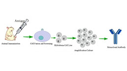 Monoclonal Antibody Customized Service
-
 Polyclonal Antibody Customized Service
-
 Protein Activity Test Experiment Service
-
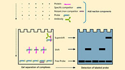 Electrophoretic Mobility Shift Assay (EMSA) Experiment Service
-
 Buffer
-
 Lentivirus Packaging Experiment Service
-
 Adenovirus Packaging Experiment Service
-
 Real Time PCR Experimental Service
-
 Spike RBD Protein (S-RBD)
-
 Protein G
-
 Protein A
Citations
- Tissue Inhibitor of Metalloproteinase-1 and -3 Improves Cardiac Function in an Ischemic Cardiomyopathy Model RatPubmed:24814095
- Fibroblast growth factor 1 ameliorates diabetic nephropathy by an anti-inflammatory mechanism.pubmed:28750927
- Characterization of fibroblast growth factor 1 in obese children and adolescentsPubmed:29991637
- Long non‑coding RNA NORAD regulates angiogenesis of human umbilical vein endothelial cells via miR‑590‑3p under hypoxic conditionsPubmed: 32323787
- Oxidative stress-induced growth inhibitor 1 in alcohol-induced liver cirrhosis34969228
- Leukocyte cell-derived chemotaxin-2 and fibroblast growth factor 21 in alcohol-induced liver cirrhosisPubmed:35070009