Active Interleukin 3 (IL3) ")

MCGF; MULTI-CSF; MCSF; M-CSF; Multiple; Multilineage-Colony-Stimulating Factor; Hematopoietic Growth Factor; P-Cell Stimulating Factor; Mast-Cell Growth Factor
- UOM
- FOB US$ 312.00 US$ 780.00 US$ 1,560.00 US$ 4,680.00 US$ 11,700.00
- Quantity
Overview
Properties
- Product No.APA076Mu61
- Organism SpeciesMus musculus (Mouse) Same name, Different species.
- ApplicationsCell culture; Activity Assays.
Research use only - DownloadInstruction Manual
- CategoryCytokineInfection immunity
- Buffer FormulationPBS, pH7.4, containing 0.01% SKL, 1mM DTT, 5% Trehalose and Proclin300.
- Traits Freeze-dried powder, Purity > 95%
- Isoelectric Point7.9
Sign into your account
Share a new citation as an author
Upload your experimental result
Review

Contact us
Please fill in the blank.
-
 Packages (Simulation)
-
 Packages (Simulation)
-
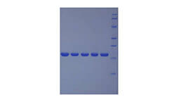
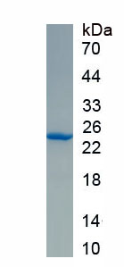 SDS-PAGE
-
 ISO9001: 2008, ISO13485: 2003 Registered
Activity test
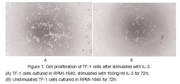
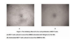
Interleukin 3 is an interleukin, a type of biological signal that can improve the body's natural response to disease as part of the immune system. It acts by binding to the interleukin 3 receptor IL-3 synergizes with other cytokines to stimulate the growth of immature progenitor cells of all lineages, and is therefore a multi-lineage colony-stimulating factor (CSF). It prevents cell death and promotes the survival of macrophages, mast cells, and megakaryocytes. To test the effect of IL-3 on cell proliferation, TF-1 cells were seeded into triplicate wells of 96-well plates at a density of 5,000 cells/well with 2% serum standard 1640 which contains various concentrations of recombinant mouse IL-3. After incubated for 72h, cells were observed by inverted microscope and cell proliferation was measured by Cell Counting Kit-8 (CCK-8). Briefly, 10 µl of CCK-8 solution was added to each well of the plate, then the absorbance at 450 nm was measured using a microplate reader after incubating the plate for 2-4 hours at 37 ℃. Proliferation of TF-1 cells after incubation with IL-3 for72h observed by inverted microscope was shown in Figure1. Cell viability was assessed by CCK-8(Cell Counting Kit-8 ) assay after incubation with recombinant IL-3 for 72h. The result was shown in Figure2. It was obvious that IL-3 significantly increased cell viability of TF-1 cells. The ED50 is 3.79 ng/ml.
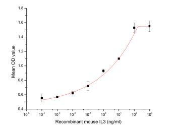
Figure 2. The dose-effect curve of IL-3 on TF-1 cells
Usage
Reconstitute in 10mM PBS (pH7.4) to a concentration of 0.1-1.0 mg/mL. Do not vortex.
Storage
Avoid repeated freeze/thaw cycles. Store at 2-8°C for one month. Aliquot and store at -80°C for 12 months.
Stability
The thermal stability is described by the loss rate. The loss rate was determined by accelerated thermal degradation test, that is, incubate the protein at 37°C for 48h, and no obvious degradation and precipitation were observed. The loss rate is less than 5% within the expiration date under appropriate storage condition.
Increment services
-
 BCA Protein Quantification Kit
-
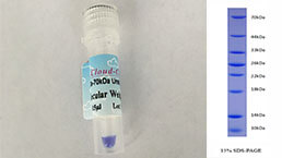 Molecular Mass Marker for Protein
-
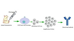 Monoclonal Antibody Customized Service
-
 Polyclonal Antibody Customized Service
-
 Protein Activity Test Experiment Service
-
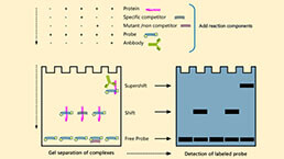 Electrophoretic Mobility Shift Assay (EMSA) Experiment Service
-
 Buffer
-
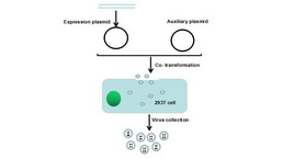 Lentivirus Packaging Experiment Service
-
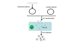 Adenovirus Packaging Experiment Service
-
 Real Time PCR Experimental Service
-
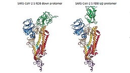 Spike RBD Protein (S-RBD)
-
 Protein G
-
 Protein A
Citations
- Effects of 900-MHz Microwave Radiation on γ-Ray-Induced Damage to Mouse Hematopoietic SystemPubMed: 20391130
- c-Kit-Positive Adipose Tissue-Derived Mesenchymal Stem Cells Promote the Growth and Angiogenesis of Breast Cancerpubmed:28573141
- Aquaporin‑4 deletion ameliorates enterovirus 71 infection in micePubmed: 32582978
- Leydig and Sertoli cell function in individuals with genital ambiguity, 46, XY karyotype, palpable gonads and normal testosterone secretion: a case-control …Pubmed:35137906