Active Mdm2 p53 Binding Protein Homolog (MDM2) ")

HDMX; hdm2; Mouse Double Minute 2,Human Homolog Of p53-Binding Protein; Oncoprotein Mdm2
Overview
Properties
- Product No.APG790Mu01
- Organism SpeciesMus musculus (Mouse) Same name, Different species.
- ApplicationsCell culture; Activity Assays.
Research use only - DownloadInstruction Manual
- CategoryTumor immunity
- Buffer Formulation20mM Tris, 150mM NaCl, pH8.0, containing 1mM EDTA, 1mM DTT, 0.01% SKL, 5% Trehalose and Proclin300.
- Traits Freeze-dried powder, Purity > 95%
- Isoelectric Point4.2
Sign into your account
Share a new citation as an author
Upload your experimental result
Review

Contact us
Please fill in the blank.
-
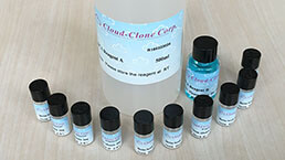
 Packages (Simulation)
-
 Packages (Simulation)
-
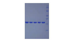
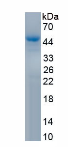 Figure. SDS-PAGE
-
 ISO9001: 2008, ISO13485: 2003 Registered
Activity test
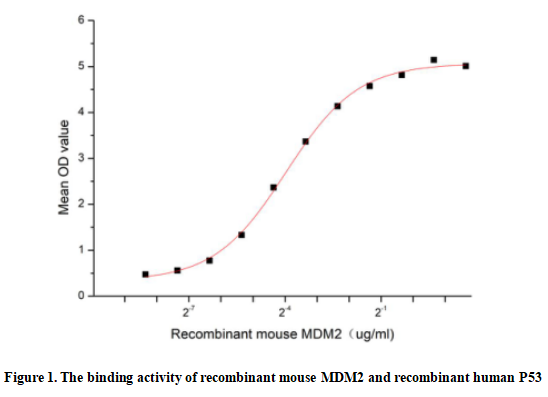
Mouse double minute 2 homolog (MDM2) also known as E3 ubiquitin-protein ligase. MdM2 is a cellular oncoprotein that recognizes the N-terminal trans-activation domain (TAD) of the p53 tumor suppressor and as an inhibitor of p53 transcriptional activation. The human homologue of this protein is sometimes called Hdm2. The p53 tumor suppressor is the key target of MDM2. It has been identified as a p53 interacting protein that represses p53 transcriptional activity and also acts as an E3 ubiquitin ligase, targeting both itself and p53 for degradation by the proteasome. MDM2 is capable of auto-polyubiquitination, and in complex with p300, a cooperating E3 ubiquitin ligase, is capable of polyubiquitinating p53. Thus a binding ELISA assay was conducted to detect the interaction of recombinant mouse MDM2 and recombinant human P53. Briefly, biotin-linked MDM2 were diluted serially in PBS, with 0.01% BSA (pH 7.4). Duplicate samples of 100μl were then transferred to P53-coated microtiter wells and incubated for 1h at 37℃. Wells were washed with PBST 3 times and incubation with Streptavidin-HRP for 30min, then wells were aspirated and washed 5 times. With the addition of substrate solution, wells were incubated 15-25 minutes at 37℃. Finally, add 50µl stop solution to the wells and read at 450nm immediately. The binding activity of MDM2 and P53 was shown in Figure 1, the EC50 for this effect is 0.064ug/mL.
Usage
Reconstitute in 20mM Tris, 150mM NaCl (pH8.0) to a concentration of 0.1-1.0 mg/mL. Do not vortex.
Storage
Avoid repeated freeze/thaw cycles. Store at 2-8°C for one month. Aliquot and store at -80°C for 12 months.
Stability
The thermal stability is described by the loss rate. The loss rate was determined by accelerated thermal degradation test, that is, incubate the protein at 37°C for 48h, and no obvious degradation and precipitation were observed. The loss rate is less than 5% within the expiration date under appropriate storage condition.
Increment services
-
 BCA Protein Quantification Kit
-
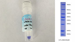 Molecular Mass Marker for Protein
-
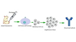 Monoclonal Antibody Customized Service
-
 Polyclonal Antibody Customized Service
-
 Protein Activity Test Experiment Service
-
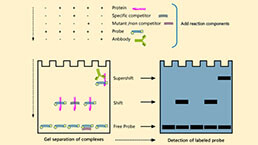 Electrophoretic Mobility Shift Assay (EMSA) Experiment Service
-
 Buffer
-
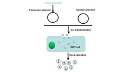 Lentivirus Packaging Experiment Service
-
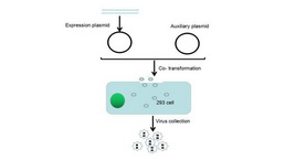 Adenovirus Packaging Experiment Service
-
 Real Time PCR Experimental Service
-
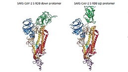 Spike RBD Protein (S-RBD)
-
 Protein G
-
 Protein A
Citations
- Inactivation of the p53–KLF4–CEBPA Axis in Acute Myeloid LeukemiaPubMed: 26408402
- Inactivation of the p53–KLF6–CEBPA Axis in Acute Myeloid LeukemiaPubMed: 26408402
- Sensitive and simultaneous surface plasmon resonance detection of free and p53-bound MDM2 proteins from human sarcomasPubmed:29637949