Active Myocilin (MYOC) ")

GLC1A; GPOA; JOAG; JOAG1; TIGR; Trabecular Meshwork Inducible Glucocorticoid Response; Myocilin 55 kDa subunit
- UOM
- FOB US$ 288.00 US$ 720.00 US$ 1,440.00 US$ 4,320.00 US$ 10,800.00
- Quantity
Overview
Properties
- Product No.APH586Mu62
- Organism SpeciesMus musculus (Mouse) Same name, Different species.
- ApplicationsCell culture; Activity Assays.
Research use only - DownloadInstruction Manual
- CategoryMetabolic pathway
- Buffer FormulationPBS, pH7.4, containing 5% Trehalose.
- Traits Freeze-dried powder, Purity > 80%
- Isoelectric Point6.1
Sign into your account
Share a new citation as an author
Upload your experimental result
Review

Contact us
Please fill in the blank.
-
 Packages (Simulation)
-
 Packages (Simulation)
-
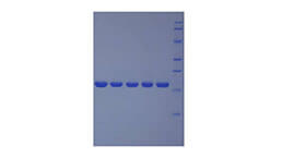
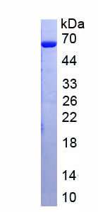 Figure. SDS-PAGE
-
 ISO9001: 2008, ISO13485: 2003 Registered
Activity test
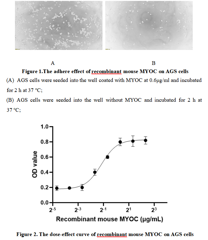
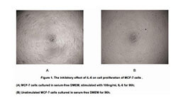
Myocilin (MYOC) is a secreted glycoprotein with a complex and multifaceted role in various biological processes, most notably associated with ocular health and the pathogenesis of glaucoma. Encoded by the MYOC gene located on chromosome 1q24.3-q25.2, this protein is primarily expressed in the trabecular meshwork (TM) of the eye, a tissue critical for regulating aqueous humor outflow and maintaining intraocular pressure (IOP). MYOC has also been detected in other tissues such as the heart, kidney, and skeletal muscle, suggesting potential extracocular functions that are still being elucidated, including roles in cell proliferation, migration, and response to mechanical stress. Research into MYOC continues to shed light on glaucoma mechanisms and may inform the development of targeted therapies for this debilitating condition. As MYOC has the function of cell adhesion, we measure the activity of recombinant mouse MYOC by the ability of the immobilized protein to support the adhesion of AGS cells. When cells are added to different concentrations of recombinant mouse MYOC coated plates, cells will adhere after 2 hour incubation at 37 ℃. The adhesion of AGS after 2 hour incubation at 37 ℃ observed by inverted microscope was shown in Figure 1. Cell adherent was in a dose dependent manner, the result was shown in Figure 2, the EC50 was 0.45 μg/ml.
Usage
Reconstitute in 10mM PBS (pH7.4) to a concentration of 0.1-1.0 mg/mL. Do not vortex.
Storage
Avoid repeated freeze/thaw cycles. Store at 2-8°C for one month. Aliquot and store at -80°C for 12 months.
Stability
The thermal stability is described by the loss rate. The loss rate was determined by accelerated thermal degradation test, that is, incubate the protein at 37°C for 48h, and no obvious degradation and precipitation were observed. The loss rate is less than 5% within the expiration date under appropriate storage condition.
Increment services
-
 BCA Protein Quantification Kit
-
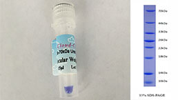 Molecular Mass Marker for Protein
-
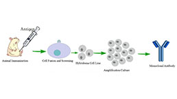 Monoclonal Antibody Customized Service
-
 Polyclonal Antibody Customized Service
-
 Protein Activity Test Experiment Service
-
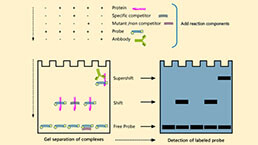 Electrophoretic Mobility Shift Assay (EMSA) Experiment Service
-
 Buffer
-
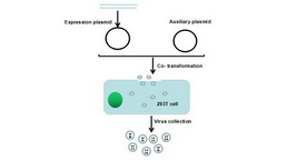 Lentivirus Packaging Experiment Service
-
 Adenovirus Packaging Experiment Service
-
 Real Time PCR Experimental Service
-
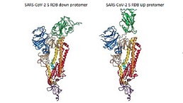 Spike RBD Protein (S-RBD)
-
 Protein G
-
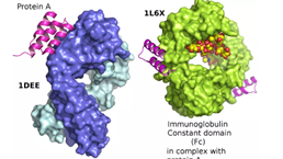 Protein A