Active Myoglobin (MYO) ")

PVALB; MB
- UOM
- FOB US$ 252.00 US$ 630.00 US$ 1,260.00 US$ 3,780.00 US$ 9,450.00
- Quantity
Overview
Properties
- Product No.APA480Hu01
- Organism SpeciesHomo sapiens (Human) Same name, Different species.
- ApplicationsCell culture; Activity Assays.
Research use only - DownloadInstruction Manual
- CategoryCardiovascular biology
- Buffer Formulation20mM Tris, 150mM NaCl, pH8.0, containing 1mM EDTA, 1mM DTT, 0.01% SKL, 5% Trehalose and Proclin300.
- Traits Freeze-dried powder, Purity > 95%
- Isoelectric Point6.9
Sign into your account
Share a new citation as an author
Upload your experimental result
Review

Contact us
Please fill in the blank.
-
 Packages (Simulation)
-
 Packages (Simulation)
-
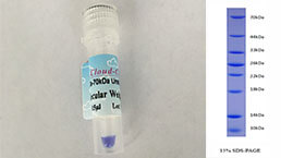
 Figure. SDS-PAGE
-
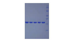
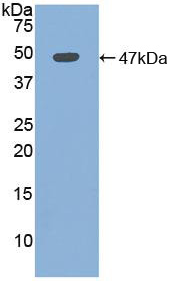 Sample: Recombinant MYO, Human;
Antibody: Rabbit Anti-Human MYO Ab (PAA480Hu01)
Figure. Western Blot
-
 ISO9001: 2008, ISO13485: 2003 Registered
Activity test
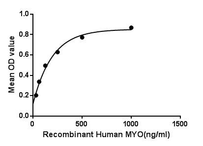
Myoglobin (symbol Mb or MB or MYO) is an iron- and oxygen-binding protein found in the muscle tissue of vertebrates in general and in almost all mammals. It is related to hemoglobin, which is the iron- and oxygen-binding protein in blood, specifically in the red blood cells. Myoglobin is found in Type I muscle, Type II A and Type II B, but most texts consider myoglobin not to be found in smooth muscle. Besides, Proteasome 26S Subunit, Non ATPase 4 (PSMD4) has been identified as an interactor of MYO, thus a binding ELISA assay was conducted to detect the interaction of recombinant human MYO and recombinant human PSMD4 Briefly, MYO were diluted serially in PBS, with 0.01% BSA (pH 7.4). Duplicate samples of 100μL were then transferred to PSMD4-coated microtiter wells and incubated for 2h at 37℃. Wells were washed with PBST and incubated for 1h with anti-MYO pAb, then aspirated and washed 3 times. After incubation with HRP labelled secondary antibody, wells were aspirated and washed 3 times. With the addition of substrate solution, wells were incubated 15-25 minutes at 37℃. Finally, add 50µL stop solution to the wells and read at 450nm immediately. The binding activity of MYO and PSMD4 was shown in Figure 1, and this effect was in a dose dependent manner.
Figure. The binding activity of MYO with PSMD4.
Usage
Reconstitute in 20mM Tris, 150mM NaCl (pH8.0) to a concentration of 0.1-1.0 mg/mL. Do not vortex.
Storage
Avoid repeated freeze/thaw cycles. Store at 2-8°C for one month. Aliquot and store at -80°C for 12 months.
Stability
The thermal stability is described by the loss rate. The loss rate was determined by accelerated thermal degradation test, that is, incubate the protein at 37°C for 48h, and no obvious degradation and precipitation were observed. The loss rate is less than 5% within the expiration date under appropriate storage condition.
Increment services
-
 BCA Protein Quantification Kit
-
 Molecular Mass Marker for Protein
-
 Monoclonal Antibody Customized Service
-
 Polyclonal Antibody Customized Service
-
 Protein Activity Test Experiment Service
-
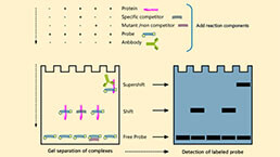 Electrophoretic Mobility Shift Assay (EMSA) Experiment Service
-
 Buffer
-
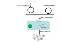 Lentivirus Packaging Experiment Service
-
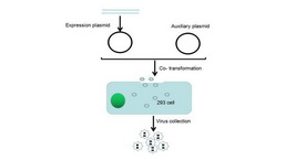 Adenovirus Packaging Experiment Service
-
 Real Time PCR Experimental Service
-
 Spike RBD Protein (S-RBD)
-
 Protein G
-
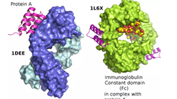 Protein A
Citations
- Acute leukocyte, cytokine and adipocytokine responses to maximal and hypertrophic resistance exercise boutsPubmed:25145982
- Transcriptional Alterations by Ischaemic Postconditioning in a Pig Infarction Model: Impact on Microvascular ProtectionPubmed: 30650650
- Electrochemical dual-aptamer-based biosensor for nonenzymatic detection of cardiac troponin I by nanohybrid electrocatalysts labeling combined with DNA …Pubmed: 30954926
- DNA nanotetrahedron linked dual-aptamer based voltammetric aptasensor for cardiac troponin I using a magnetic metal-organic framework as a labelPubmed: 31123904
- Emitter–Quencher Pair of Single Atomic Co Sites and Monolayer Titanium Carbide MXenes for Luminol Chemiluminescent Reactions34914377
- A rapid fluorescent aptasensor for point-of-care detection of C-reactive proteinPubmed:35714415