Active Perforin 1 (PRF1) ")

FLH2; HPLH2; P1; PFP; Pore Forming Protein; Cytolysin; Lymphocyte pore-forming protein
- UOM
- FOB US$ 314.00 US$ 785.00 US$ 1,570.00 US$ 4,710.00 US$ 11,775.00
- Quantity
Overview
Properties
- Product No.APB317Mu03
- Organism SpeciesMus musculus (Mouse) Same name, Different species.
- ApplicationsCell culture; Activity Assays.
Research use only - DownloadInstruction Manual
- CategoryImmune molecule
- Buffer Formulation20mM Tris, 150mM NaCl, pH8.0, containing 0.01% SKL, 5% Trehalose.
- Traits Freeze-dried powder, Purity > 80%
- Isoelectric Point8.0
Sign into your account
Share a new citation as an author
Upload your experimental result
Review

Contact us
Please fill in the blank.
-
 Packages (Simulation)
-
 Packages (Simulation)
-
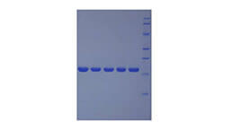
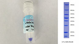
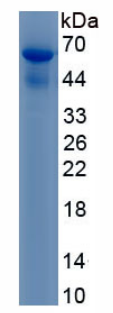 Figure. SDS-PAGE
-
 ISO9001: 2008, ISO13485: 2003 Registered
Activity test
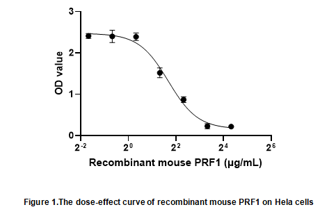
Perforin 1 (PRF1) is a cytolytic protein found in the granules of Cytotoxic T lymphocytes (CTLs) and NK cells. Upon degranulation, perforin inserts itself into the target cell's plasma membrane, forming a pore. The lytic membrane-inserting part of perforin is the MACPF domain. This region shares homology with cholesterol-dependent cytolysins from Gram-positive bacteria. It has been demonstrated that PRF1 causes the lysis of abnormal body cells and the elimination of virus-infected cells and tumors.To test the effect of PRF1 on cell apoptosis,Hela cells were seeded into triplicate wells of 96-well plates and allowed to attach, replaced with various concentrations of recombinant mouse PRF1. After incubated for 72h, cells were observed by inverted microscope and cell proliferation was measured by Cell Counting Kit-8 (CCK-8). Briefly, 10 µl of CCK-8 solution was added to each well of the plate, then the absorbance at 450 nm was measured using a microplate reader after incubating the plate for 1-4 hours at 37 ℃. Cell viability was assessed by CCK-8 assay after incubation with recombinant mouse PRF1 for 72h. The result was shown in Figure 1. It was obvious that PRF1 significantly decreased cell viability of Hela cells. The ED50 of recombinant mouse PRF1 is 3.205 μg/ml.
Usage
Reconstitute in 20mM Tris, 150mM NaCl (pH8.0) to a concentration of 0.1-1.0 mg/mL. Do not vortex.
Storage
Avoid repeated freeze/thaw cycles. Store at 2-8°C for one month. Aliquot and store at -80°C for 12 months.
Stability
The thermal stability is described by the loss rate. The loss rate was determined by accelerated thermal degradation test, that is, incubate the protein at 37°C for 48h, and no obvious degradation and precipitation were observed. The loss rate is less than 5% within the expiration date under appropriate storage condition.
Increment services
-
 BCA Protein Quantification Kit
-
 Molecular Mass Marker for Protein
-
 Monoclonal Antibody Customized Service
-
 Polyclonal Antibody Customized Service
-
 Protein Activity Test Experiment Service
-
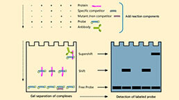 Electrophoretic Mobility Shift Assay (EMSA) Experiment Service
-
 Buffer
-
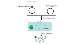 Lentivirus Packaging Experiment Service
-
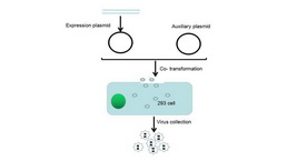 Adenovirus Packaging Experiment Service
-
 Real Time PCR Experimental Service
-
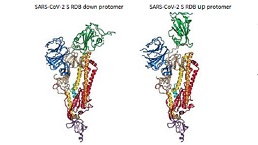 Spike RBD Protein (S-RBD)
-
 Protein G
-
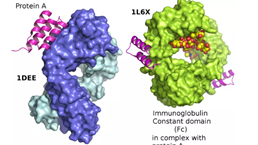 Protein A
Citations
- Granzyme B as a diagnostic marker of tuberculosis in patients with and without HIV coinfectionPubmed:26915636
- The immunoregulatory effects of CD8 T-cell–derived perforin on diet-induced nonalcoholic steatohepatitisPubmed: 30951375
- A novel Granzyme B nanoparticle delivery system simulates immune cell functions for suppression of solid tumorsPubmed: 31695790
- CD317 mediates immunocytolysis resistance by RICH2/cytoskeleton-dependent membrane protectionPubmed: 33223223
- Dual-function chimeric antigen receptor T cells targeting c-Met and PD-1 exhibit potent anti-tumor efficacy in solid tumorsPubmed: 32772342
- Combined treatment with epigenetic agents enhances anti-tumor activity of T cells by upregulating the ACRBP expression in hepatocellular carcinoma34377237
- Cancer-cell stiffening via cholesterol depletion enhances adoptive T-cell immunotherapy34873307
- Single-cell immune profiling reveals functional diversity of T cells in tuberculous pleural effusionPubmed:35061012