Active Platelet Derived Growth Factor D (PDGFD) ")

PDGF-D; IEGF; SCDGF-B; Spinal Cord Derived Growth Factor B; Iris-expressed growth factor; Platelet-derived growth factor D, receptor-binding form/latent form
- UOM
- FOB US$ 314.00 US$ 785.00 US$ 1,570.00 US$ 4,710.00 US$ 11,775.00
- Quantity
Overview
Properties
- Product No.APC919Hu01
- Organism SpeciesHomo sapiens (Human) Same name, Different species.
- ApplicationsCell culture; Activity Assays.
Research use only - DownloadInstruction Manual
- CategoryCytokine
- Buffer Formulation20mM Tris, 150mM NaCl, pH8.0, containing 1mM EDTA, 1mM DTT, 0.01% SKL, 5% Trehalose and Proclin300.
- Traits Freeze-dried powder, Purity > 90%
- Isoelectric Point8.4
Sign into your account
Share a new citation as an author
Upload your experimental result
Review

Contact us
Please fill in the blank.
-
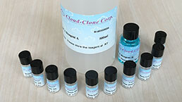
 Packages (Simulation)
-
 Packages (Simulation)
-
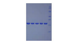
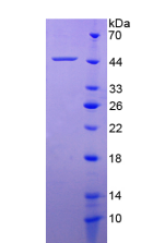 Figure. SDS-PAGE
-
 ISO9001: 2008, ISO13485: 2003 Registered
Activity test
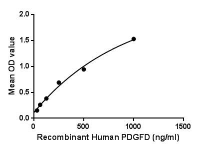
Platelet-derived growth factor D (PDGFD) is a protein that in humans is encoded by the PDGFD gene. The protein encoded by this gene is a member of the platelet-derived growth factor family. PDGF plays a significant role in blood vessel formation, the growth of blood vessels from already-existing blood vessel tissue, mitogenesis. PDGF also plays a role in embryonic development, cell proliferation, cell migration, and angiogenesis. Besides, Macrophage Erythroblast Attacher (MAEA) has been identified as an interactor of PDGFD, thus a binding ELISA assay was conducted to detect the interaction of recombinant human PDGFD and recombinant human MAEA. Briefly, PDGFD were diluted serially in PBS, with 0.01% BSA (pH 7.4). Duplicate samples of 100μL were then transferred to MAEA-coated microtiter wells and incubated for 2h at 37℃. Wells were washed with PBST and incubated for 1h with anti-PDGFD pAb, then aspirated and washed 3 times. After incubation with HRP labelled secondary antibody, wells were aspirated and washed 3 times. With the addition of substrate solution, wells were
incubated 15-25 minutes at 37℃. Finally, add 50µL stop solution to the wells and read at 450nm immediately. The binding activity of PDGFD and MAEA was shown in Figure 1, and this effect was in a dose dependent manner.
Figure. The binding activity of PDGFD with MAEA.
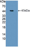
Figure. Western Blot
Usage
Reconstitute in 20mM Tris, 150mM NaCl (PH8.0) to a concentration of 0.1-1.0 mg/mL. Do not vortex.
Storage
Avoid repeated freeze/thaw cycles. Store at 2-8°C for one month. Aliquot and store at -80°C for 12 months.
Stability
The thermal stability is described by the loss rate. The loss rate was determined by accelerated thermal degradation test, that is, incubate the protein at 37°C for 48h, and no obvious degradation and precipitation were observed. The loss rate is less than 5% within the expiration date under appropriate storage condition.
Increment services
-
 BCA Protein Quantification Kit
-
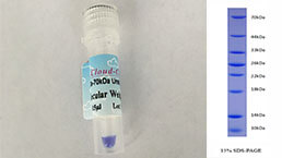 Molecular Mass Marker for Protein
-
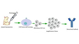 Monoclonal Antibody Customized Service
-
 Polyclonal Antibody Customized Service
-
 Protein Activity Test Experiment Service
-
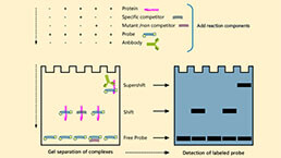 Electrophoretic Mobility Shift Assay (EMSA) Experiment Service
-
 Buffer
-
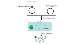 Lentivirus Packaging Experiment Service
-
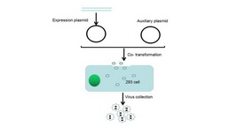 Adenovirus Packaging Experiment Service
-
 Real Time PCR Experimental Service
-
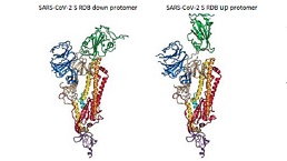 Spike RBD Protein (S-RBD)
-
 Protein G
-
 Protein A
Citations
- In vitro gene silencing effect of chitosan/shRNA PDGF-D nanoparticles in breast cancerDOI: 10.12991/mpj.2017.21
- Transforming Growth Factor-β3 Regulates Adipocyte Number in Subcutaneous White Adipose TissuePubmed: 30332637