Active Transforming Growth Factor Beta 2 (TGFb2) ")

TGF-B2; TGF-Beta2; G-TSF; LAP; Cetermin; Polyergin; Latency-associated peptide; BSC-1 cell growth inhibitor; Glioblastoma-derived T-cell suppressor factor
- UOM
- FOB US$ 330.00 US$ 824.00 US$ 1,648.00 US$ 4,944.00 US$ 12,360.00
- Quantity
Overview
Properties
- Product No.APA218Hu01
- Organism SpeciesHomo sapiens (Human) Same name, Different species.
- ApplicationsCell culture; Activity Assays.
Research use only - DownloadInstruction Manual
- CategoryCytokineInfection immunity
- Buffer FormulationPBS, pH7.4, containing 0.01% SKL, 5% Trehalose.
- Traits Freeze-dried powder, Purity > 90%
- Isoelectric Point8.9
Sign into your account
Share a new citation as an author
Upload your experimental result
Review

Contact us
Please fill in the blank.
-
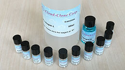
 Packages (Simulation)
-
 Packages (Simulation)
-
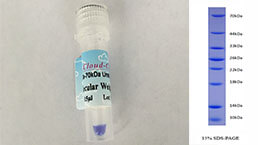
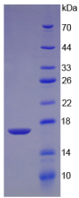 Figure. SDS-PAGE
-
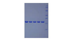
 WB Image
-
 ISO9001: 2008, ISO13485: 2003 Registered
Activity test

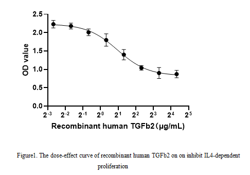
Transforming Growth Factor Beta 2 (TGFB2) belongs to TGF-beta family. As a secreted protein and a cytokine, TGFB2 plays a role in the formation of blood vessels, the regulation of muscle tissue and body fat development, wound healing, and immune system function. Besides, TGFB2 regulates cell proliferation, differentiation, adhesion, migration by transducing the signal through transmembrane receptors type I and type II (TGFBR1 and TGFBR2). To test the effect of TGFb2 on inhibit IL4-dependent proliferation, CTLL-2 cells were seeded into triplicate wells of 96-well plates, the medium was 2% serum RPMI-1640 including various concentrations of recombinant human TGFb2. After incubated for 72h, cells were observed by inverted microscope and cell proliferation was measured by Cell Counting Kit-8 (CCK-8). Briefly, 10 µl of CCK-8 solution was added to each well of the plate, then the absorbance at 450 nm was measured. Cell viability was assessed by CCK-8 assay. The result was shown in Figure1. It was obvious that TGFB2 significantly decreased cell viability of CTLL-2 cells. The ED50 of recombinant human TGFB2 is 1.95μg/mL.
Usage
Reconstitute in 10mM PBS (pH7.4) to a concentration of 0.1-1.0 mg/mL. Do not vortex.
Storage
Avoid repeated freeze/thaw cycles. Store at 2-8°C for one month. Aliquot and store at -80°C for 12 months.
Stability
The thermal stability is described by the loss rate. The loss rate was determined by accelerated thermal degradation test, that is, incubate the protein at 37°C for 48h, and no obvious degradation and precipitation were observed. The loss rate is less than 5% within the expiration date under appropriate storage condition.
Increment services
-
 BCA Protein Quantification Kit
-
 Molecular Mass Marker for Protein
-
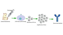 Monoclonal Antibody Customized Service
-
 Polyclonal Antibody Customized Service
-
 Protein Activity Test Experiment Service
-
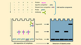 Electrophoretic Mobility Shift Assay (EMSA) Experiment Service
-
 Buffer
-
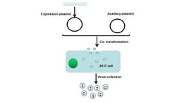 Lentivirus Packaging Experiment Service
-
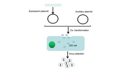 Adenovirus Packaging Experiment Service
-
 Real Time PCR Experimental Service
-
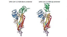 Spike RBD Protein (S-RBD)
-
 Protein G
-
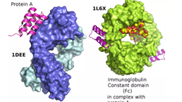 Protein A
Citations
- EVALUATION OF PHYSICO-CHEMICAL PROPERTIES OF COLOSTRUM SUPPLEMENTED DAHIIjfans: Source
- The immunomodulating effect of seminal plasma on T cellsPubMed: 25799173
- Studies on quality attributes of skimmed colostrum powderP-ISSN: 2349–8528
- Angiocrine signals regulate quiescence and therapy resistance in bone metastasisPubmed: 31292293
- Association of CASC18/miR-20a-3p/TGFB2 ceRNA axis with occult lymph node metastasis in tongue squamous cell carcinoma34362313
- Effect of convection and microwave heating on the retention of bioactive components in human milk34896952