Transmission Electron Microscopy (TEM) Experiment Service
Instruction manual
FOR IN VITRO AND RESEARCH USE ONLY
NOT FOR USE IN CLINICAL DIAGNOSTIC PROCEDURES
First Edition (Revised on April, 2016)
Information
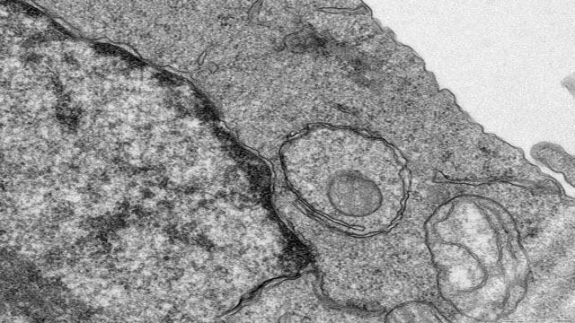
Transmission Electron Microscope (TEM) can see fine structure less than 0.2um which can not see in the optical microscope, these structures are called submicron structures or ultrastructures. In order to see these structures, it is should to select light source with a shorter wavelength to improve the resolution of the microscope. The resolution of TEM can reach 0.2nm.
TEM is usually used in materials science, biology. Because electrons are easily scattered or absorbed by objects and the penetration is low, so the density, thickness, of the sample will affect the final image quality. It should be prepare thin slice(50-100nm) sample for TEM.
Service Content
Observation for Cell samples, microbial samples, and tissue samples
Service Procedure
1. Prepare test samples(Customer or our company provide).
2. Perform TEM according to customer requirements, provide to customer test image.
Imaging Instrument
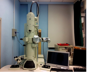
JEM-1400 120kV TEM


Customer Providing
1. The treated samples, or fresh tissue samples
2. It should be determine the sample material location, minimize the mechanical damage such as traction, contusion and compression. Tissue volume is generally not more than 1mm×1mm×1mm. Quickly put the samples into the electron microscope fix solution at 4 ℃ for 2-4hours.


