


The versatility of the IFN family: From basic immune response to precise disease intervention
The versatility of the IFN family: From basic immune response to precise disease intervention
1. Introduction to interferon family
The interferon (IFN) family is a group of signaling proteins that play a crucial role in the body's antiviral immunity. These cytokines are induced when cells are infected by viruses. These cytokines establish a defense line by interfering with virus replication, activating immune cells, and enhancing the antiviral state of host cells. Due to their broad antiviral, antiproliferative, and immunomodulatory activities, they have become important biological agents for clinical treatment of viral hepatitis, certain malignant tumors, and multiple sclerosis and other diseases.
1.1 Distribution of the IFN Family
The IFN family is mainly classified into three categories based on receptor specificity, sequence homology, and biological functions. Type I IFNs, including IFNα, IFNβ, IFNε, IFNκ, and IFNω, among which IFNα and IFNβ are the most well-characterized and deeply studied. Type I IFNs are produced by various cell types, such as monocytes, macrophages, B cells, T cells, platelets, epithelial cells, endothelial cells, and tumor cells, and play a central role in the innate immunity of the body, directly interfering with viral replication and establishing a "antiviral state" in cells. Type II IFNs are represented only by IFNγ, mainly produced by activated immune cells, including T lymphocytes, B lymphocytes, natural killer (NK) cells, and natural killer T (NKT) cells, focusing on activating macrophages, enhancing antigen presentation to regulate acquired immunity. Type III IFNs consist of four members: IFNλ1, IFNλ2, IFNλ3, and IFNλ4, mainly produced by epithelial cells and plasmacytoid dendritic cells, providing a first-line antiviral defense at the mucosal barrier.
1.2 Structure of the IFN Family
Each IFN consists of six secondary structural elements, denoted as A-F. Among them, the helices A, C, D, and F form an antiparallel four-helix bundle. The secondary structures of the ring elements B and E are more diverse, ranging from additional helices to extended segments, which are arranged closely along the edge of the four-helix bundle. The α helix of type I IFN is long and straight, basically parallel to each other. Unlike type I interferons, type III interferons are composed of shorter and more twisted helices, forming a more compact bundle-like structure. Unlike the monomeric forms of type I and type III interferons, IFNγ adopts an intercalated dimeric structure, where the helices E and F of one chain interchange with the corresponding helices of the other subunit in the dimer. This highlights the different mechanisms by which each interferon family regulates biological activity: IFNγ functions through receptor homodimerization, while the monomeric forms of type I and type III interferons rely on variable contacts between the interferon and the receptor.
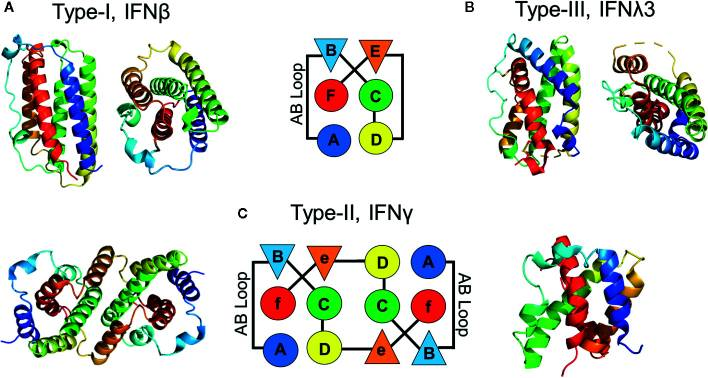
Fig.1 The structure of IFN family (Sourced from Front Immunol[1])
1.3 IFN Family Signal Transduction
When interferons bind to specific receptors on the cell membrane, they activate the JAK kinase coupled to the receptors, thereby causing phosphorylation modifications to the signal transduction and transcriptional activation factors (STAT); these activated STAT proteins form homodimers or heterodimers and combine with DNA-binding proteins and other components to form the interferon-stimulated gene factor 3 (ISGF3) complex, rapidly translocating to the cell nucleus and binding to the interferon-stimulated response elements in the genome to initiate the transcriptional expression of hundreds of interferon-stimulated genes. These gene products work together to exert core functions such as antiviral replication inhibition, suppression of cell proliferation, and regulation of immune responses, jointly establishing a cellular antiviral state and maintaining the immune homeostasis of the organism.
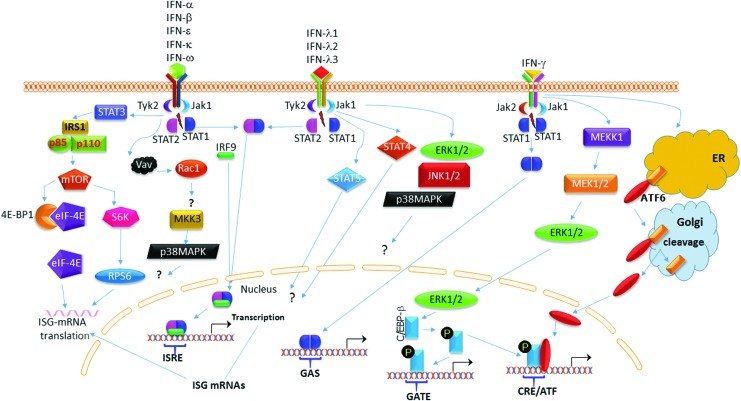
Fig.2 IFN signaling pathway (Sourced from J Interferon Cytokine Res[2])
2. IFN Family and Cancer
IFN, as a crucial immune regulatory factor, has evolved from a simple anti-proliferative application to a complex immunological combination therapy stage in cancer research. The loss of tumor suppressor transcription factor Elf5 in triple-negative breast cancer (TNBC) cells activates endogenous IFNγ signaling, promoting tumor progression and metastasis[3]. IL1RA mediates type I IFN response in oral squamous cell carcinoma (OSCC) and inhibits the malignant progression of OSCC[4]. Low-dose IFNα significantly enhances the anti-tumor properties of valproic acid, and their combined use may become an innovative option for the treatment of advanced prostate cancer[5]. Antibodies targeting PD-L1 with IFNα can improve tumor targeting and antigen presentation, and overcome resistance to checkpoint blockade therapy[6]. Using mesenchymal stem cells as an effective targeting vector, 5FU prodrug and IFNβ are delivered to the tumor site, thereby increasing local treatment concentration and significantly inhibiting the growth of peritoneal cancer[7]. Yes-associated protein (YAP) mediates the IFNγ-promoted tumor effect through its nuclear phase separation, and disrupting YAP phase separation can reduce tumor growth and make tumor cells more sensitive to PD-1 therapy[8]. The upregulation of IFNλ reprograms macrophages to an anti-tumor state, enhances phagocytosis and secretion of inflammatory cytokines, and activates adaptive immunity to inhibit the progression of bladder cancer[9]. These advancements are continuously reshaping the position of IFN in precision oncology, transforming it from a classic immune stimulator to a promising multifunctional anti-cancer platform.
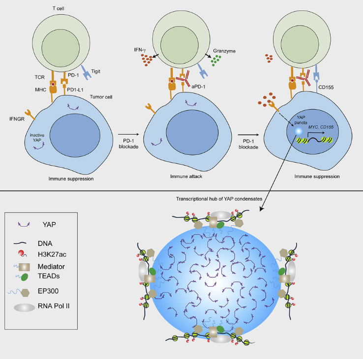
Fig.3 IFNγ induces tumor resistance to anti-PD-1 immunotherapy by promoting YAP phase separation (Sourced from Mol Cell[8])
3. IFN Family and Autoimmune diseases
IFN plays a crucial role in the immune system, and the regulation of the IFN pathway has become a new direction for the treatment of autoimmune diseases. IFN is considered a key molecule in the pathogenesis of systemic lupus erythematosus (SLE), and high levels of circulating type I, II, and III IFN are associated with significant clinical features of SLE, as well as a higher tendency for SLE-related nephritis, arthritis, skin inflammation, and other severe manifestations[10, 11]. In patients with systemic sclerosis (SSc), IFN type I characteristics were observed in the early stage of the disease before obvious skin fibrosis[12]. IFNλ1 may participate in the progression of SLE's kidney diseases and arthritis by stimulating the secretion of chemokines IP-10, MIG, and IL-8, and is related to disease activity[13]. Rheumatoid arthritis (RA) patients have elevated serum IFNλ1 levels and are associated with knee joint diseases[14]. Anifrolumab is a monoclonal antibody targeting IFNAR1 and has been proven to reduce the disease activity, glucocorticoid dosage, and severity of skin diseases in SLE patients[15]. Sifalimumab is a monoclonal antibody targeting IFNα, and in patients with moderate to severe active SLE treated with Sifalimumab, SLE response indicators improve, and specific disease activity in skin and joint tissues is also reduced[16]. With a deeper understanding of the pathogenesis and its impact on immune dysregulation and clinical heterogeneity, targeted precision medicine targeting IFN has become a very promising treatment strategy in autoimmune diseases.
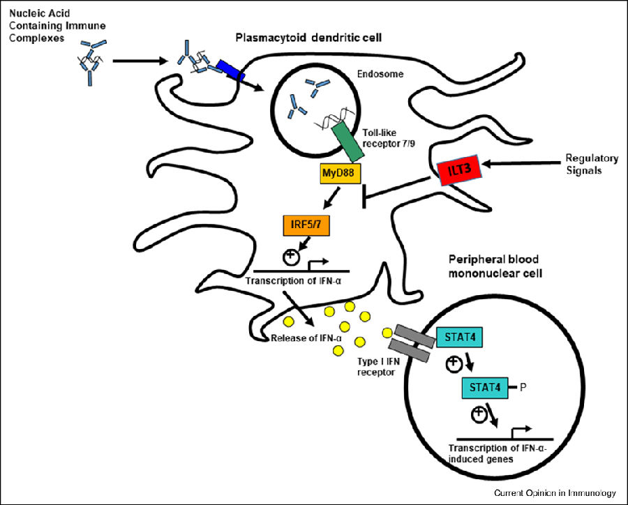
Fig.4 Schematic diagram of type I IFN pathway regulation in SLE (Sourced from Curr Opin Immunol[10])
4. IFN Family and Central nervous system diseases
A large amount of evidence indicates that the IFN signal is closely related to neuroinflammation within the central nervous system (CNS) and plays a crucial role in the pathogenesis of various neurological and neurodegenerative diseases. By treating Parkinson's disease (PD) mouse models with IFNAR1 monoclonal antibodies, the neuroinflammation and dopaminergic neuron cell death were reduced, confirming the potential of targeting the type I IFN pathway for neuroprotection[17]. Strong and amyloid-beta (Aβ)-pathology-dependent type I IFN activation was detected in microglia and other cell types, and blocking the type I IFN signal could rescue memory and synaptic defects, and reduce inflammation and neuropathy[18]. The type I IFN-stimulated genes (ISGs) were significantly upregulated in patients with amyotrophic lateral sclerosis (ALS), and IFN pathway inhibitors treatment reduced IFN response markers, slowed disease progression, and prolonged the survival of ALS mice[19]. In the experimental autoimmune encephalomyelitis mouse model, IFNλ promoted disease maintenance and axonal damage by maintaining effector Th1 cells in the central nervous system[20]. IFNγ stimulation enhanced the processing of α-synuclein preformed fibrils in microglia, promoted the generation of neuroprotective metabolites, and partially maintained the energy supply of dopaminergic neurons[21]. With the deepening understanding of the role of interferons in the neuro-immune intersection field, targeted interference based on interferons is expected to bring new breakthroughs in neurological diseases.
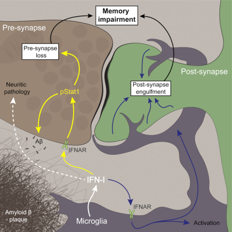
Fig.5 Type I IFN signaling synergistically promotes memory impairment associated with Aβ plaques (Sourced from Immunity[18])
5. IFN Family and Central nervous system diseases
IFNβ enhances the adhesion between macrophages and endothelial cells, and promotes the attraction of white blood cells to the sites prone to atherosclerosis in a chemokine-dependent manner[22]. IFNβ treatment accelerates lesion formation in a mouse atherosclerosis model and increases the accumulation of macrophages in plaques. The increase in type I interferon activity may lead to elevated expression of DNA sensors genes in colonic epithelial cells of ulcerative colitis (UC) and JAK-dependent inflammatory cell death[23]. Overexpression of IFNβ1 can alleviate fat inflammation caused by obesity and regulate adipose tissue hypertrophy[24]. These effects are related to the inhibition of weight gain and restoration of glucose homeostasis. IFN-α-2b prevents weight gain and dyslipidemia induced by a high-fat diet through mechanisms involving fatty acid oxidation and cholesterol reduction[25]. IFNγ inhibits the proliferation of CD4+ T cells that produce pathogenic IL-17A by interacting with receptors on the pulmonary parenchymal cell population, and plays a protective role in idiopathic pulmonary fibrosis[26]. IFNλ promotes the proliferation of regulatory T cells expressing Foxp3 and inhibits the generation of IL-5 and IL-13 in vitro, playing an important role in the pathogenesis of allergic asthma[27]. Therefore, a deeper understanding of the correlation between interferon signaling and different diseases can provide important basis for the development of new diagnostic and therapeutic strategies targeting interferon signaling.
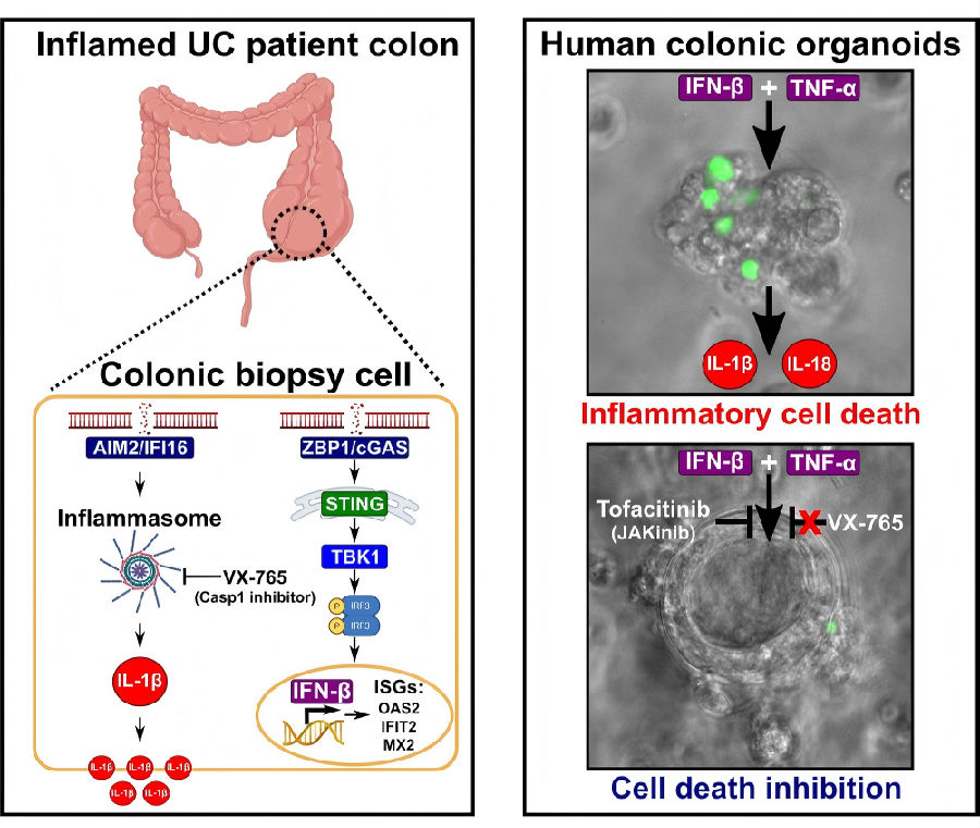
Fig.6 Type I IFN signaling increases in UC and induces JAK dependent inflammatory cell death
(Sourced from Am J Physiol Gastrointest Liver Physiol[23])
Cloud-Clone supports scientific research and provides relevant detection reagent products for a wide range of scientific researchers. The core product numbers of the relevant targets are as follows:
Target | core product No. | Target | core product No. | Target | core product No. |
CXCR3 | A625 | IFNa21 | G966 | IRF8 | B776 |
GBP1 | E637 | IFNa4 | A175 | IRF9 | H780 |
GBP2 | E636 | IFNa5 | G975 | ITaC | C071 |
IFI16 | B900 | IFNa7 | G973 | JAK1 | C551 |
IFI30 | E762 | IFNa8 | G972 | JAK2 | F494 |
IFI35 | L605 | IFNa9 | S760 | MAP2K1 | D559 |
IFI44 | P030 | IFNb | A222 | MAP2K2 | D562 |
IFIH1 | L608 | IFNe | D175 | MAP2K4 | MKK4 |
IFIT1 | L609 | IFNg | A049 | MAPK11 | B435 |
IFITM2 | H829 | IFNgR1 | B491 | MAPK12 | D577 |
IFITM3 | H830 | IFNgR2 | L627 | MAPK13 | D578 |
IFNa | A033 | IFNk | D176 | MAPK14 | B206 |
IFNa/bR1 | B425 | IFNt | B862 | MIg | B928 |
IFNa/bR2 | E171 | IFNw | B569 | PDK1 | C718 |
IFNa10 | G971 | IP10 | A371 | PIK3Cb | J829 |
IFNa11 | S090 | IRF1 | B564 | PIK3Cd | J832 |
IFNa13 | G970 | IRF2 | C180 | RPS6Kb1 | L979 |
IFNa14 | G969 | IRF3 | B589 | STAT1 | B740 |
IFNa16 | G968 | IRF4 | B755 | STAT2 | B796 |
IFNa17 | G967 | IRF5 | B598 | TYK2 | B595 |
IFNa2 | A179 | IRF6 | B958 |
For more scientific research reagents, please visit the official website of Cloud-Clone:http://www.cloud-clone.com/
References
[1]Walter MR. The Role of Structure in the Biology of Interferon Signaling. Front Immunol. 2020;11:606489.
[2]Nallar SC, Kalvakolanu DV. Interferons, signal transduction pathways, and the central nervous system. J Interferon Cytokine Res. 2014;34(8):559-576.
[3]Singh S, Kumar S, Srivastava RK, et al. Loss of ELF5-FBXW7 stabilizes IFNGR1 to promote the growth and metastasis of triple-negative breast cancer through interferon-γ signalling. Nat Cell Biol. 2020;22(5):591-602.
[4]Ding Y, Shan Y, Gu J, Yi J, Sun Z. IL1RA inhibits the progression of oral squamous cell carcinoma by mediating type Ⅰ interferon response. Transl Oncol. 2025;58:102428.
[5]Hudak L, Tezeeh P, Wedel S, et al. Low dosed interferon alpha augments the anti-tumor potential of histone deacetylase inhibition on prostate cancer cell growth and invasion. Prostate. 2012;72(16):1719-1735.
[6]Liang Y, Tang H, Guo J, et al. Targeting IFNα to tumor by anti-PD-L1 creates feedforward antitumor responses to overcome checkpoint blockade resistance. Nat Commun. 2018;9(1):4586.
[7]Ho YK, Woo JY, Loke KM, Deng LW, Too HP. Enhanced anti-tumor efficacy with multi-transgene armed mesenchymal stem cells for treating peritoneal carcinomatosis. J Transl Med. 2024;22(1):463.
[8]Yu M, Peng Z, Qin M, et al. Interferon-γ induces tumor resistance to anti-PD-1 immunotherapy by promoting YAP phase separation. Mol Cell. 2021;81(6):1216-1230.e9.
[9]Wang B, Zhou B, Chen J, et al. Type III interferon inhibits bladder cancer progression by reprogramming macrophage-mediated phagocytosis and orchestrating effective immune responses. J Immunother Cancer. 2024;12(4):e007808.
[10]Postal M, Vivaldo JF, Fernandez-Ruiz R, Paredes JL, Appenzeller S, Niewold TB. Type I interferon in the pathogenesis of systemic lupus erythematosus. Curr Opin Immunol. 2020;67:87-94.
[11]Oke V, Gunnarsson I, Dorschner J, et al. High levels of circulating interferons type I, type II and type III associate with distinct clinical features of active systemic lupus erythematosus. Arthritis Res Ther. 2019;21(1):107.
[12]Brkic Z, van Bon L, Cossu M, et al. The interferon type I signature is present in systemic sclerosis before overt fibrosis and might contribute to its pathogenesis through high BAFF gene expression and high collagen synthesis. Ann Rheum Dis. 2016;75(8):1567-1573.
[13]Wu Q, Yang Q, Lourenco E, Sun H, Zhang Y. Interferon-lambda1 induces peripheral blood mononuclear cell-derived chemokines secretion in patients with systemic lupus erythematosus: its correlation with disease activity. Arthritis Res Ther. 2011;13(3):R88.
[14]Wu Q, Yang Q, Sun H, Li M, Zhang Y, La Cava A. Serum IFN-λ1 is abnormally elevated in rheumatoid arthritis patients. Autoimmunity. 2013;46(1):40-43.
[15]Furie R, Khamashta M, Merrill JT, et al. Anifrolumab, an Anti-Interferon-α Receptor Monoclonal Antibody, in Moderate-to-Severe Systemic Lupus Erythematosus. Arthritis Rheumatol. 2017;69(2):376-386.
[16]Khamashta M, Merrill JT, Werth VP, et al. Sifalimumab, an anti-interferon-α monoclonal antibody, in moderate to severe systemic lupus erythematosus: a randomised, double-blind, placebo-controlled study. Ann Rheum Dis. 2016;75(11):1909-1916.
[17]Main BS, Zhang M, Brody KM, et al. Type-1 interferons contribute to the neuroinflammatory response and disease progression of the MPTP mouse model of Parkinson's disease. Glia. 2016;64(9):1590-1604.
[18]Roy ER, Chiu G, Li S, et al. Concerted type I interferon signaling in microglia and neural cells promotes memory impairment associated with amyloid β plaques. Immunity. 2022;55(5):879-894.e6.
[19]Carletta O, Perfetto C, Rifai OM, et al. Genotype-specific interferon signatures in amyotrophic lateral sclerosis relate to disease severity. Brain. 2026;149(2):489-501.
[20]Manivasagam S, Williams JL, Vollmer LL, et al. Targeting IFN-λ Signaling Promotes Recovery from Central Nervous System Autoimmunity. J Immunol. 2022;208(6):1341-1351.
[21]Niskanen J, Hakosalo V, Hämäläinen W, et al. IFNγ alters the aberrant phenotype of α-synuclein-treated microglia reducing the detrimental impact of their secretome on dopaminergic neurons. Neuroscience. 2026;603:201-213.
[22]Goossens P, Gijbels MJ, Zernecke A, et al. Myeloid type I interferon signaling promotes atherosclerosis by stimulating macrophage recruitment to lesions. Cell Metab. 2010;12(2):142-153.
[23]Flood P, Fanning A, Woznicki JA, et al. DNA sensor-associated type I interferon signaling is increased in ulcerative colitis and induces JAK-dependent inflammatory cell death in colonic organoids. Am J Physiol Gastrointest Liver Physiol. 2022;323(5):G439-G460.
[24]Alsaggar M, Mills M, Liu D. Interferon beta overexpression attenuates adipose tissue inflammation and high-fat diet-induced obesity and maintains glucose homeostasis. Gene Ther. 2017;24(1):60-66.
[25]Quiroga AD, Comanzo CG, Heit Barbini FJ, et al. IFN-α-2b treatment protects against diet-induced obesity and alleviates non-alcoholic fatty liver disease in mice. Toxicol Appl Pharmacol. 2019;379:114650.
[26]Mauermann N, Burian J, von Garnier C, et al. Interferon-gamma regulates idiopathic pneumonia syndrome, a Th17+CD4+ T-cell-mediated graft-versus-host disease. Am J Respir Crit Care Med. 2008;178(4):379-388.
[27]Koch S, Finotto S. Role of Interferon-λ in Allergic Asthma. J Innate Immun. 2015;7(3):224-230.