- SPF
Mouse Model for Cerebral Ischemia (CI) ")

Brain Ischemia; Cerebrovascular Ischemia; MCAO
- UOM
- FOB US$ 300.00
- Quantity
Overview
Properties
- Product No.DSI523Mu01
- Organism SpeciesMus musculus (Mouse) Same name, Different species.
- ApplicationsDiesase model
Research use only - Downloadn/a
- Category
- Prototype SpeciesHuman
- SourceIschemia-Reperfusion with MCAO
- Model Animal StrainsBalb/c Mice(SPF level), male, healthy, body weight 25g~30g
- Modeling GroupingRandomly divided into groups: Control group, Model group, Positive drug group and Test drug group, 15 mice per group.
- Modeling Period24 hours, 3 days or 7 days
Sign into your account
Share a new citation as an author
Upload your experimental result
Review

Contact us
Please fill in the blank.
-
 Packages (Simulation)
-
 Packages (Simulation)
-
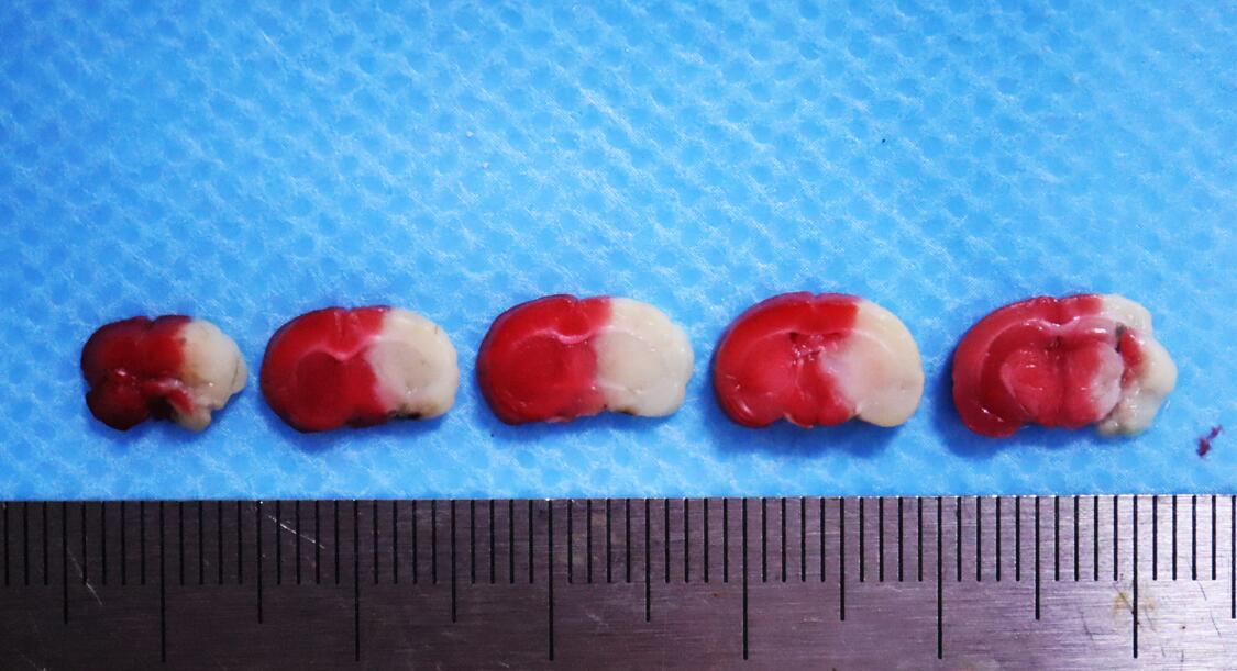 Fig.TTC staining for modeling of mice brain ischemia-reperfusion
-
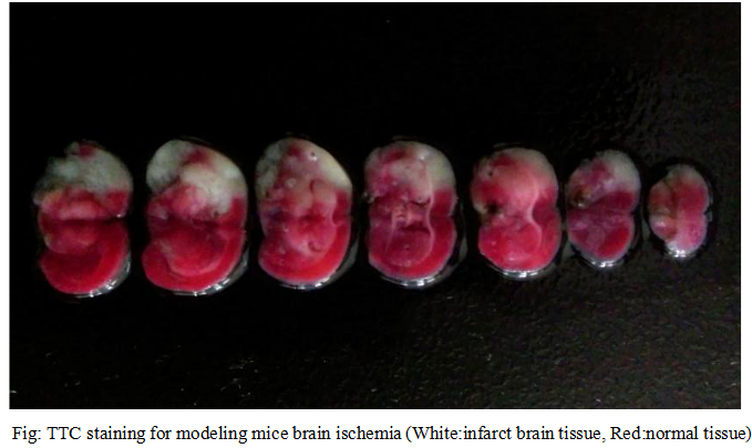 Fig.TTC staining for modeling of mice brain ischemia-reperfusion
-
 ISO9001: 2008, ISO13485: 2003 Registered
Modeling Method
1. The mice were anesthetized, the neck skin was prepared, the anus temperature probe was inserted, and the body temperature was kept at 37±0.5℃..
2. Incision of the middle of the neck, exposure the right common carotid artery, internal carotid artery and external carotid artery. Using 7-0 silk (at 2mm distance from the common carotid artery bifurcation ) to ligate the external carotid artery at the far end of the heart, pierces another 7-0 silk in the external carotid artery , and hit a slipknot near the bifurcation of the common carotid artery.
3. Use arterial clamps to clamp the common carotid artery. Make a small cut on the external carotid artery (1.5 mm distance from bifurcation of common carotid artery ), a root tip treated 0.18 mm diameter nylon line from the cut insertion into the internal carotid artery, and inward into the middle cerebral artery, nylon line insertion depth distance carotid artery bifurcation about 9±1mm.
4. Ischemia 60 mins and pull the suture, a 7-0 silk ligates artery proximal, 5-0 silk suture wounds on the neck, povidone iodine disinfection wound, put mice in a heating pad, and feed on constant temperature raising box after mice awaked.
5. 24h after operation, neurological function score is evaluated, and then the mice are anesthetized, and the brains are stained with TTC and pathological staining.
Model evaluation
1.Neurological dysfunction score
Longa and Bederson's 5 point systems,make a score on mice awaked from anaesthesia 24 hours later.
0 points: no nerve injury symptoms
1 points can not fully extend the forepaw
2 points: Turn to the opposite side
3 points:Tip to the opposite side
4 points: can not walk spontaneously, loss of consciousness
2.TTC staining
Take the brain and store at -20℃, 1% TTC (W/V), 37℃ water bath for TTC dissolved, the frozen brain tissue slices, placed in 10ml TTC solution, 37℃, 10min. Normal brain tissue staining is bright red, and the infarct area is pale.
Pathological results
Take the brain, 4% poly formaldehyde solution fixed, after dehydration of sucrose solution, the OCT embedded to make the frozen section (slice 10um), Nissl staining and the staining resluts are used for the evaluation of infarct size.
Cytokines level
Statistical analysis
SPSS software is used for statistical analysis, measurement data to mean ± standard deviation (x ±s), using t test and single factor analysis of variance for group comparison , P<0.05 indicates there was a significant difference, P<0.01 indicates there are very significant differences.
Giveaways

Increment services
-
 Tissue/Sections Customized Service
-
 Serums Customized Service
-
 Immunohistochemistry (IHC) Experiment Service
-
 Small Animal In Vivo Imaging Experiment Service
-
 Small Animal Micro CT Imaging Experiment Service
-
 Small Animal MRI Imaging Experiment Service
-
 Small Animal Ultrasound Imaging Experiment Service
-
 Transmission Electron Microscopy (TEM) Experiment Service
-
 Scanning Electron Microscope (SEM) Experiment Service
-
 Learning and Memory Behavioral Experiment Service
-
 Anxiety and Depression Behavioral Experiment Service
-
 Drug Addiction Behavioral Experiment Service
-
 Pain Behavioral Experiment Service
-
 Neuropsychiatric Disorder Behavioral Experiment Service
-
 Fatigue Behavioral Experiment Service
-
 Nitric Oxide Assay Kit (A012)
-
 Nitric Oxide Assay Kit (A013-2)
-
 Total Anti-Oxidative Capability Assay Kit(A015-2)
-
 Total Anti-Oxidative Capability Assay Kit (A015-1)
-
 Superoxide Dismutase Assay Kit
-
 Fructose Assay Kit (A085)
-
 Citric Acid Assay Kit (A128 )
-
 Catalase Assay Kit
-
 Malondialdehyde Assay Kit
-
 Glutathione S-Transferase Assay Kit
-
 Microscale Reduced Glutathione assay kit
-
 Glutathione Reductase Activity Coefficient Assay Kit
-
 Angiotensin Converting Enzyme Kit
-
 Glutathione Peroxidase (GSH-PX) Assay Kit
-
 Cloud-Clone Multiplex assay kits