Mouse Model for Hypoxic Ischemic Encephalopathy (HIE) 

HIBD; NE; Hypoxic-ischemic brain damage; Neonatal encephalopathy
- UOM
- FOB US$
- Quantity
Overview
Properties
- Product No.DSI685Mu02
- Organism SpeciesMus musculus (Mouse) Same name, Different species.
- ApplicationsDisease Model
Research use only - Downloadn/a
- CategoryCerebral and nervous systems
- Prototype SpeciesHuman
- SourceInduced by hypoxia
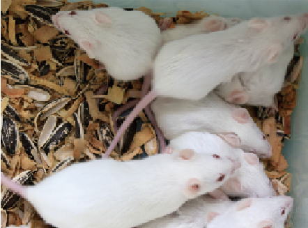
- Model Animal StrainsBalb/c Mice(SPF level), male, healthy, 6~8 weeks.
- Modeling GroupingRandomly divided into groups: Control group, Model group, Positive drug group and Test drug group, 15 mice per group
- Modeling Period4~6w
Sign into your account
Share a new citation as an author
Upload your experimental result
Review

Contact us
Please fill in the blank.
Modeling Method
Place mouse in an visual isobaric chamber, pour into hypoxia gas 5L/min (8%O2 and 92%N2). The treatment last for 90min every time, 1-3 times for a week, treatment period were 3 weeks.
Model evaluation
Morris water maze:
At the age of 12 weeks, morris water maze for all groups. The experiment is divided into place navigation test and spatial probe test.
1. Place navigation test: Rats were trained for 5 days, 4 times a day, 30min for each interval. Record the time it takes rats to find the platform from 4 entry points, i.e. incubation. The average score of the four incubation as the final result of the day entered the final statistics.
2. Spatial probe test: 6th days of the test, remove platform, from the furthest end of the platform into the water. Record the swimming track of rats within 30s, observe and analyse the residence time in the target quadrant and times to pass through the platform.
Pathological results
After behavior detection, remove brain in each group, peel the epencephal on ice, fixed in 4% paraformaldehyde for pathological examination.
1. IHC
Observation of Aβ-amyloid plaques staining in hippocampus and cortex. Under the light microscope, count and analyze the number of positive plaque at the same site of each group for 6 fields.
2. Thiolain S staining
Detection of Aβ-amyloid plaques (green fluorescence) in hippocampus and cortex by using Thiolain S staining. Under the fluorescence microscope, count and analyze the number of positive plaque at the same site of each group for 6 fields.
Cytokines level
Statistical analysis
SPSS software is used for statistical analysis, measurement data to mean ± standard deviation (x ±s), using t test and single factor analysis of variance for group comparison , P<0.05 indicates there was a significant difference, P<0.01 indicates there are very significant differences.
Giveaways
Increment services
-
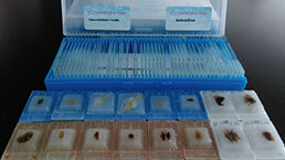 Tissue/Sections Customized Service
-
 Serums Customized Service
-
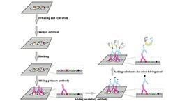 Immunohistochemistry (IHC) Experiment Service
-
 Small Animal In Vivo Imaging Experiment Service
-
 Small Animal Micro CT Imaging Experiment Service
-
 Small Animal MRI Imaging Experiment Service
-
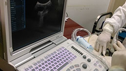 Small Animal Ultrasound Imaging Experiment Service
-
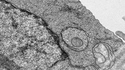 Transmission Electron Microscopy (TEM) Experiment Service
-
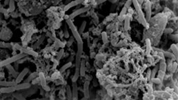 Scanning Electron Microscope (SEM) Experiment Service
-
 Learning and Memory Behavioral Experiment Service
-
 Anxiety and Depression Behavioral Experiment Service
-
 Drug Addiction Behavioral Experiment Service
-
 Pain Behavioral Experiment Service
-
 Neuropsychiatric Disorder Behavioral Experiment Service
-
 Fatigue Behavioral Experiment Service
-
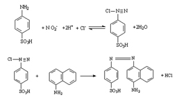 Nitric Oxide Assay Kit (A012)
-
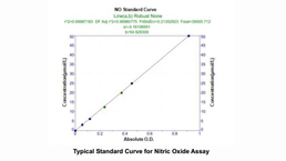 Nitric Oxide Assay Kit (A013-2)
-
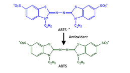 Total Anti-Oxidative Capability Assay Kit(A015-2)
-
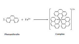 Total Anti-Oxidative Capability Assay Kit (A015-1)
-
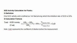 Superoxide Dismutase Assay Kit
-
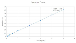 Fructose Assay Kit (A085)
-
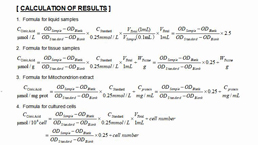 Citric Acid Assay Kit (A128 )
-
 Catalase Assay Kit
-
 Malondialdehyde Assay Kit
-
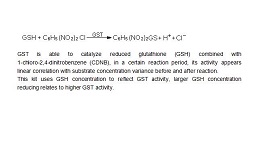 Glutathione S-Transferase Assay Kit
-
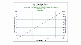 Microscale Reduced Glutathione assay kit
-
 Glutathione Reductase Activity Coefficient Assay Kit
-
 Angiotensin Converting Enzyme Kit
-
 Glutathione Peroxidase (GSH-PX) Assay Kit
-
 Cloud-Clone Multiplex assay kits


