Mouse Model for Osteoporosis (OP) ")

- UOM
- FOB US$ 220.00
- Quantity
Overview
Properties
- Product No.DSI534Mu01
- Organism SpeciesMus musculus (Mouse) Same name, Different species.
- ApplicationsDisease Model
Research use only - Downloadn/a
- Category
- Prototype SpeciesHuman
- SourceInduced by ovariectomy
- Model Animal StrainsC57BL/6 mice (SPF level), healthy, female not pregnant, week age:6W~8W, body weight 18g~20g
- Modeling GroupingRandomly divided into groups: Control group, Model group, Positive drug group and Test drug group, 15 mice per group.
- Modeling Period12 weeks
Sign into your account
Share a new citation as an author
Upload your experimental result
Review

Contact us
Please fill in the blank.
-
 Packages (Simulation)
-
 Packages (Simulation)
-
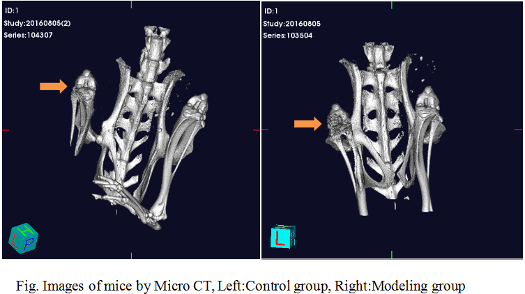 Fig. Images of mice by Micro CT
-
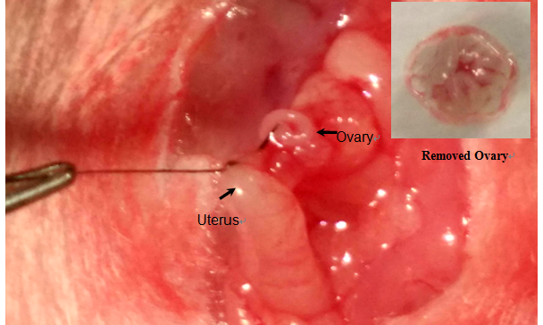 Fig. The operation of mice ovariectomy
-
 ISO9001: 2008, ISO13485: 2003 Registered
Modeling Method
1. 3% sodium pentobarbital (40mg/kg)intraperitoneal injection of anesthesia, remove the abdominal hair, skin disinfection.
2. Cut off the skin and the muscle layer in the middle of the abdomen on the sterile condition.
3. Separate the uterus and remove the ovaries, and then suture the wound, after the mice awake, put them back to clean cage, observe the state and death of mice, and make records.
4. 3 days after surgery, injecte 40 thousand units of penicillin to prevent infection for each mouse.
5. For control group ,only remove about 1g fat around the ovary, while the ovaries are retained, and the other operation are the same.
6. X-ray measurement of bone mineral density( BMD) on 12W, and intraperitoneal injection of anesthesia, get blood from eyeball, 4℃, 3000r/min,10min, separate the upper serum and stored at 80℃ for preservation of the refrigerator spare. Take the tibia of mice,and make pathological examination.
Model evaluation
1. The changes of body weight, estradiol and bone mineral density in ovariectomized mice
In the ovariectomized group, the body weight increased rapidly, the activity ability decreased, the hair color is dull and gray, while the weight of the control group increased slowly, the hair color is bright, and the activity is normal. Estradiol levels and BMD of femur in the ovariectomized group are significantly decreased.
Pathological results
From each mouse tibia specimens, placed in 10% neutral formalin fixed buffer solution (pH=7.2-7.4,4℃) fixed 24-48h. Then the 10%EDTA-2 Na buffer solution (pH = 7.4, 4℃) decalcification, every 5-7d to renew the decalcifying fluid; demineralized completely, dehydrated in graded ethanol and xylene transparent, longitudinal paraffin embedded, 5um sections, HE staining.
In the control group, the trabecular bone of the tibia is concentrated and interwoven into a network; the number of trabecular bone in the model group is significantly reduced, and the trabecular bone becomes thin.
Cytokines level
Some studies have found that the lack of estrogen will lead to spontaneous reaction of the body. The expression of IL-6, IL-8, IL-1 and TNF-α in the serum of ovariectomized is increased, which could be detected by ELISA method.
Statistical analysis
SPSS software is used for statistical analysis, measurement data to mean ± standard deviation (x ±s), using t test and single factor analysis of variance for group comparison , P<0.05 indicates there was a significant difference, P<0.01 indicates there are very significant differences.
Giveaways

Increment services
-
 Tissue/Sections Customized Service
-
 Serums Customized Service
-
 Immunohistochemistry (IHC) Experiment Service
-
 Small Animal In Vivo Imaging Experiment Service
-
 Small Animal Micro CT Imaging Experiment Service
-
 Small Animal MRI Imaging Experiment Service
-
 Small Animal Ultrasound Imaging Experiment Service
-
 Transmission Electron Microscopy (TEM) Experiment Service
-
 Scanning Electron Microscope (SEM) Experiment Service
-
 Learning and Memory Behavioral Experiment Service
-
 Anxiety and Depression Behavioral Experiment Service
-
 Drug Addiction Behavioral Experiment Service
-
 Pain Behavioral Experiment Service
-
 Neuropsychiatric Disorder Behavioral Experiment Service
-
 Fatigue Behavioral Experiment Service
-
 Nitric Oxide Assay Kit (A012)
-
 Nitric Oxide Assay Kit (A013-2)
-
 Total Anti-Oxidative Capability Assay Kit(A015-2)
-
 Total Anti-Oxidative Capability Assay Kit (A015-1)
-
 Superoxide Dismutase Assay Kit
-
 Fructose Assay Kit (A085)
-
 Citric Acid Assay Kit (A128 )
-
 Catalase Assay Kit
-
 Malondialdehyde Assay Kit
-
 Glutathione S-Transferase Assay Kit
-
 Microscale Reduced Glutathione assay kit
-
 Glutathione Reductase Activity Coefficient Assay Kit
-
 Angiotensin Converting Enzyme Kit
-
 Glutathione Peroxidase (GSH-PX) Assay Kit
-
 Cloud-Clone Multiplex assay kits