Mouse Model for Pulmonary Fibrosis (PF) ")

Lung Fibrosis
- UOM
- FOB US$ 200.00
- Quantity
Overview
Properties
- Product No.DSI518Mu01
- Organism SpeciesMus musculus (Mouse) Same name, Different species.
- Applicationsn/a
Research use only - Downloadn/a
- Category
- Prototype SpeciesHuman
- Sourceinduce by Bleomycin
- Model Animal StrainsBalb/c Mice(SPF), healthy, male, age: 8~10weeks, body weight:20g~25g.
- Modeling GroupingRandomly divided into six group: Control group, Model group, Positive drug group and Test drug group.
- Modeling Period4-6 weeks
Sign into your account
Share a new citation as an author
Upload your experimental result
Review

Contact us
Please fill in the blank.
-
 Packages (Simulation)
-
 Packages (Simulation)
-
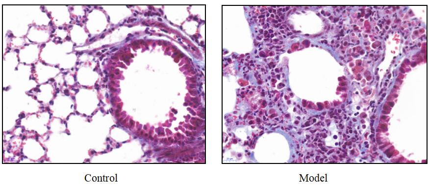 Fig.MASSON staining for mouse lung
-
 ISO9001: 2008, ISO13485: 2003 Registered
Modeling Method
Modeling method:
SPF C57/BL6 female mice, weight about 18~20g. The model was modeled by tracheal drip bleomycin.
The anesthetized mice were fixed in supine position on the experimental table. After the neck hair was removed, the skin was cut and the trachea was exposed layer by layer. A 1 mL syringe was pierced into the trachea through the gap between the two tracheal cartilage rings and toward the heart, and bleomycin was injected 5mg/kg/L (the control group was injected with the same volume of normal saline) if there was no resistance.
Model evaluation
Development time of pulmonary fibrosis model:On the 7th day after administration, most of the lung tissues showed severe alveolitis, with a large number of neutrophils infiltrating in the alveolar cavity and interstitium, some alveolar cavities destroyed or disappeared, and fibroblasts and capillaries hyperplasia in the lung septum, which were significantly different from normal lung tissues;On the 14th day after administration, pulmonary fibrosis began to form. Macrophages, neutrophils and other inflammatory cells decreased significantly, fibroblasts increased, alveolar septum thickened significantly, collagen deposition; On day 28 after administration, most of the mice developed diffuse pulmonary interstitial fibrosis, in which the pulmonary interstitial was replaced by collagen fibers and fibroblasts, alveolar wall was destroyed, and pulmonary bullae formed, but inflammatory cell infiltration was still visible.
Pathological results
Cytokines level
Statistical analysis
SPSS software is used for statistical analysis, measurement data to mean ± standard deviation (x ±s), using t test and single factor analysis of variance for group comparison , P<0.05 indicates there was a significant difference, P<0.01 indicates there are very significant differences.
Giveaways

Increment services
-
 Tissue/Sections Customized Service
-
 Serums Customized Service
-
 Immunohistochemistry (IHC) Experiment Service
-
 Small Animal In Vivo Imaging Experiment Service
-
 Small Animal Micro CT Imaging Experiment Service
-
 Small Animal MRI Imaging Experiment Service
-
 Small Animal Ultrasound Imaging Experiment Service
-
 Transmission Electron Microscopy (TEM) Experiment Service
-
 Scanning Electron Microscope (SEM) Experiment Service
-
 Learning and Memory Behavioral Experiment Service
-
 Anxiety and Depression Behavioral Experiment Service
-
 Drug Addiction Behavioral Experiment Service
-
 Pain Behavioral Experiment Service
-
 Neuropsychiatric Disorder Behavioral Experiment Service
-
 Fatigue Behavioral Experiment Service
-
 Nitric Oxide Assay Kit (A012)
-
 Nitric Oxide Assay Kit (A013-2)
-
 Total Anti-Oxidative Capability Assay Kit(A015-2)
-
 Total Anti-Oxidative Capability Assay Kit (A015-1)
-
 Superoxide Dismutase Assay Kit
-
 Fructose Assay Kit (A085)
-
 Citric Acid Assay Kit (A128 )
-
 Catalase Assay Kit
-
 Malondialdehyde Assay Kit
-
 Glutathione S-Transferase Assay Kit
-
 Microscale Reduced Glutathione assay kit
-
 Glutathione Reductase Activity Coefficient Assay Kit
-
 Angiotensin Converting Enzyme Kit
-
 Glutathione Peroxidase (GSH-PX) Assay Kit
-
 Cloud-Clone Multiplex assay kits