Rat Model for Chronic Pancreatitis (CP) ")

- UOM
- FOB US$ 180.00
- Quantity
Overview
Properties
- Product No.DSI754Ra01
- Organism SpeciesRattus norvegicus (Rat) Same name, Different species.
- ApplicationsDisease Model
Research use only - Downloadn/a
- CategoryDigestive systemEndocrine system
- Prototype SpeciesHuman
- SourceInduced by dibutyltin dichloride (DBTC)
- Model Animal StrainsSD Rats(SPF), healthy, male and female, body weight 180g~200g.
- Modeling GroupingRandomly divided into six group: Control group, Model group, Positive drug group and Test drug group.
- Modeling Period4-6 weeks
Sign into your account
Share a new citation as an author
Upload your experimental result
Review

Contact us
Please fill in the blank.
-
 Packages (Simulation)
-
 Packages (Simulation)
-
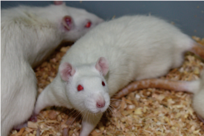 Fig. Chronic Pancreatitis (CP) rats
-
 ISO9001: 2008, ISO13485: 2003 Registered
Modeling Method
SD rats were randomly divided into 3 groups. These 3 groups were modeled for 4 weeks, 6 weeks and 8 weeks. 12 hours before the experiment, provide water but not food.
DBTC dissolved in 100% alcohol, then mix with glycerol (the ratio is 1:2:3). Rats fixed were induced by intravenous injection of 200ul mixture (DBTC 8mg/kg).
Model evaluation
Detection of food intake and body mass:
Record average food intake and body mass. Rats in model group were depressed, lacklustre, reduced appetite , and more frequent in defecation with dilute stools.
Pathological results
Observation of pancreatic pathology
Normal sections were stained with HE. Refer to Matsumura and other scoring systems for grading.
Changes in pancreatic pathology: At day 1, there was a slight swelling in the pancreas for DBTC model group. At day 3, swelling and hyperemia were obvious, pancreatic duct dilated slightly, and hydatoncus formed initially. At day 7, hydatoncus enlarged gradually, pancreas adheres to the surrounding tissue. At day 14, some giant hydatoncus formed, the pancreas began to atrophy and nodules formed. At day 28, pancreas atrophied, the color turned white, the nodule was more obvious than before, and some pancreatic cysts were complicated and infected.
Under the microscope, pancreatic edema, interstitial enlargement, massive inflammatory cell infiltrationand and partial acinar necrosis were observed in the model group at day 1; acinar atrophy, fibroblast proliferation, periacinar fibrous tissue formation and inflammatory cell infiltration were observed at day 3; fibrosis around the acinus were observed at day 7; with the exception of inflammatory cell infiltration and periacinar fibrosis, interlobular and periductal fibrosis was observed at day 14; pancreatic fibrosis was observed at day 28.
Cytokines level
Determination of amylase, protein and hydroxyproline
Expression determination of amylase, protein and hydroxyproline: Determination of hydroxyproline by Alkaline Hydrolysis. Determination of protein content by BCA method. Determination of pancreatic amylase by ELISA.
Expression changes of protein, amylase and hydroxyproline: In the model group, pancreatic protein and amylase decrease gradually and hydroxyproline increased gradually.
Statistical analysis
SPSS software is used for statistical analysis, measurement data to mean ± standard deviation (x ±s), using t test and single factor analysis of variance for group comparison , P<0.05 indicates there was a significant difference, P<0.01 indicates there are very significant differences.
Giveaways

Increment services
-
 Tissue/Sections Customized Service
-
 Serums Customized Service
-
 Immunohistochemistry (IHC) Experiment Service
-
 Small Animal In Vivo Imaging Experiment Service
-
 Small Animal Micro CT Imaging Experiment Service
-
 Small Animal MRI Imaging Experiment Service
-
 Small Animal Ultrasound Imaging Experiment Service
-
 Transmission Electron Microscopy (TEM) Experiment Service
-
 Scanning Electron Microscope (SEM) Experiment Service
-
 Learning and Memory Behavioral Experiment Service
-
 Anxiety and Depression Behavioral Experiment Service
-
 Drug Addiction Behavioral Experiment Service
-
 Pain Behavioral Experiment Service
-
 Neuropsychiatric Disorder Behavioral Experiment Service
-
 Fatigue Behavioral Experiment Service
-
 Nitric Oxide Assay Kit (A012)
-
 Nitric Oxide Assay Kit (A013-2)
-
 Total Anti-Oxidative Capability Assay Kit(A015-2)
-
 Total Anti-Oxidative Capability Assay Kit (A015-1)
-
 Superoxide Dismutase Assay Kit
-
 Fructose Assay Kit (A085)
-
 Citric Acid Assay Kit (A128 )
-
 Catalase Assay Kit
-
 Malondialdehyde Assay Kit
-
 Glutathione S-Transferase Assay Kit
-
 Microscale Reduced Glutathione assay kit
-
 Glutathione Reductase Activity Coefficient Assay Kit
-
 Angiotensin Converting Enzyme Kit
-
 Glutathione Peroxidase (GSH-PX) Assay Kit
-
 Cloud-Clone Multiplex assay kits