Rat Model for Intervertebral Disc Degeneration (DDD) ")

- UOM
- FOB US$ 400.00
- Quantity
Overview
Properties
- Product No.DSI739Ra01
- Organism SpeciesRattus norvegicus (Rat) Same name, Different species.
- ApplicationsDisease Model
Research use only - Downloadn/a
- CategorySkeletal, articular and cutaneous systems
- Prototype SpeciesHuman
- SourceInduced by surgical method
- Model Animal StrainsSD rats (SPF class), Male, 6~8W, 200~250g
- Modeling GroupingRandomly divided into six group: Control group, Model group, Positive drug group and Test drug group
- Modeling Period16~20 weeks
Sign into your account
Share a new citation as an author
Upload your experimental result
Review

Contact us
Please fill in the blank.
-
 Packages (Simulation)
-
 Packages (Simulation)
-
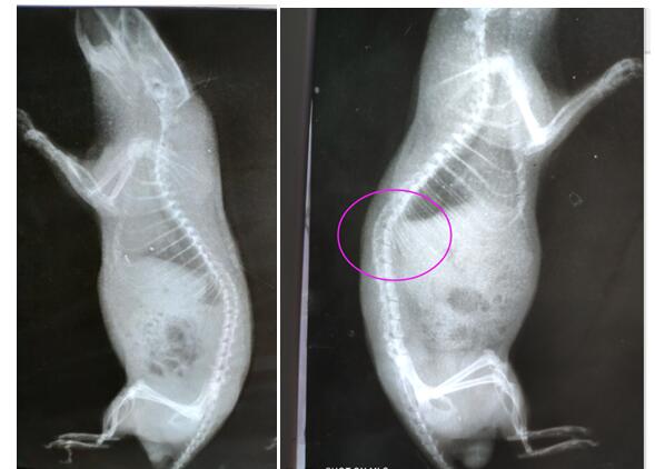 Fig. X ray image of rats,left:control,right:model
-
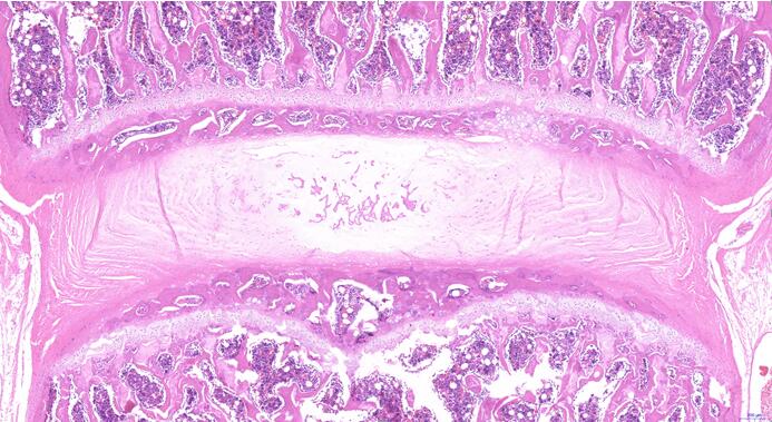 Fig. HE staining of intervertebral disc for model rat
-
 ISO9001: 2008, ISO13485: 2003 Registered
Modeling Method
After the rats were anesthetized, the posterior median incision was performed,longitudinal incision of the skin and subcutaneous tissue from the atlanto occipital joint to the second thoracic spinous process ranged from 2 to 2.5 cm, blunt free neck muscles to expose the cervical spine sufficiently. From the inside to the outside layer in order to completely cut off 2 ~ 7 cervical interspinous ligament and supraspinous ligament, deep neck atlantoaxial splenius cervicis, longissimus and iliocostalis cervicis and semispinalis muscle, superficial platysma, neck, head and neck trapezius rhomboideus, hemostasis after suture on both sides of sacrospinalis and skin. Continuous 3d injection of penicillin to prevent infection after operation.
Model evaluation
Three months later, all rats were taken anteroposterior and lateral X-ray + lumbar spine
Each segment was dyed with a solid green color (4 segments);
Materials: The whole disc and the upper and lower parts of the vertebral body should also have, the disc unit refers to the disc tissue + upper and lower endplate cartilage + upper and lower vertebral body;
Staining: take the most central part of the disc and make a coronal section (the coronal section is from the ventral side to the dorsal side). Staining should be done on the basis of the entire disc unit. The main observation is the endplate cartilage between the vertebral body and the intervertebral disc.
Pathological results
Cytokines level
Statistical analysis
SPSS software is used for statistical analysis, measurement data to mean ± standard deviation (x ±s), using t test and single factor analysis of variance for group comparison , P<0.05 indicates there was a significant difference, P<0.01 indicates there are very significant differences.
Giveaways

Increment services
-
 Tissue/Sections Customized Service
-
 Serums Customized Service
-
 Immunohistochemistry (IHC) Experiment Service
-
 Small Animal In Vivo Imaging Experiment Service
-
 Small Animal Micro CT Imaging Experiment Service
-
 Small Animal MRI Imaging Experiment Service
-
 Small Animal Ultrasound Imaging Experiment Service
-
 Transmission Electron Microscopy (TEM) Experiment Service
-
 Scanning Electron Microscope (SEM) Experiment Service
-
 Learning and Memory Behavioral Experiment Service
-
 Anxiety and Depression Behavioral Experiment Service
-
 Drug Addiction Behavioral Experiment Service
-
 Pain Behavioral Experiment Service
-
 Neuropsychiatric Disorder Behavioral Experiment Service
-
 Fatigue Behavioral Experiment Service
-
 Nitric Oxide Assay Kit (A012)
-
 Nitric Oxide Assay Kit (A013-2)
-
 Total Anti-Oxidative Capability Assay Kit(A015-2)
-
 Total Anti-Oxidative Capability Assay Kit (A015-1)
-
 Superoxide Dismutase Assay Kit
-
 Fructose Assay Kit (A085)
-
 Citric Acid Assay Kit (A128 )
-
 Catalase Assay Kit
-
 Malondialdehyde Assay Kit
-
 Glutathione S-Transferase Assay Kit
-
 Microscale Reduced Glutathione assay kit
-
 Glutathione Reductase Activity Coefficient Assay Kit
-
 Angiotensin Converting Enzyme Kit
-
 Glutathione Peroxidase (GSH-PX) Assay Kit
-
 Cloud-Clone Multiplex assay kits