Rat Model for Retinal Ischemia Reperfusion Injury (RIRI) ")

- UOM
- FOB US$ 240.00
- Quantity
Overview
Properties
- Product No.DSI846Ra01
- Organism SpeciesRattus norvegicus (Rat) Same name, Different species.
- ApplicationsDisease Model
Research use only - Downloadn/a
- Category
- Prototype SpeciesHuman
- SourceAnterior chamber pressure perfusion
- Model Animal StrainsSD Rats(SPF), healthy, male, 6~8W ,body weight 180g~200g.
- Modeling Grouping1.Randomly divided into six group: Control group, Model group, Positive drug group and Tested drug group.
- Modeling Period1w
Sign into your account
Share a new citation as an author
Upload your experimental result
Review

Contact us
Please fill in the blank.
-
 Packages (Simulation)
-
 Packages (Simulation)
-
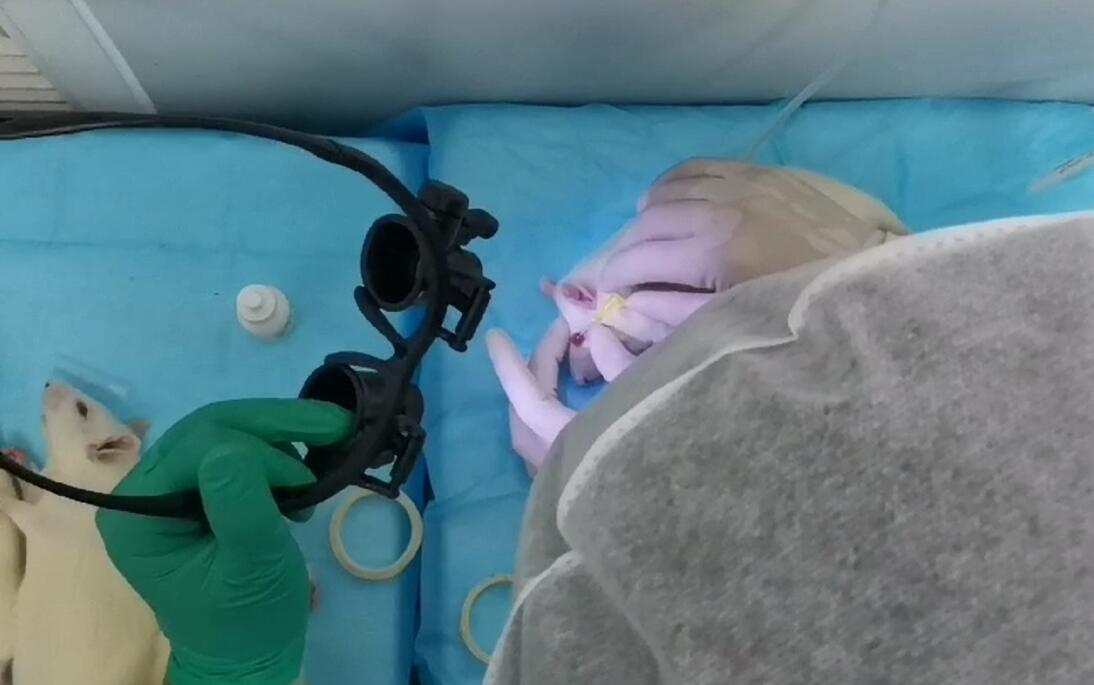 Fig.The operation of Anterior chamber pressure perfusion
-
 ISO9001: 2008, ISO13485: 2003 Registered
Modeling Method
1. Prepare for anesthesia: accurately weighing the weight of the rats and intraperitoneal injection of anesthesia rats;
2. The rats were placed on the operating table, and the right eye of the rats was disinfected with active iodine solution, and kept warm at 37℃.
3. Apply the eye anesthetic evenly to the eyeball.When there was no response that the cotton swab was touched to the eyeball of the rats, the needle of disposable intravenous infusion device was inserted into the middle and back vitreous from the sclera to stop the needle, and the eyeball was observed to change from red to colorless by sliding the flow regulator to the appropriate flow.Retinal ischemia began at this point, and the needle was removed 45 minutes later.During ischemia, care should be taken to stabilize the needle and continue anesthesia to prevent the needle from falling out or moving.
Model evaluation
After 72h of modeling, the animals were sacrificed, the retinal morphology was observed by HE staining, and the changes of Bcl-2 protein and Bax protein in retinal cells were detected by immunohistochemistry.
Results :HE staining showed that the retinal structure of normal control rats was clear and the cells of each layer were closely arranged. In the ischemia-reperfusion group, the retina is highly edematous with loss and disordered distribution of ganglion cells. A large number of Bcl-2 positive cells were observed in the retina of the normal control group, but decreased significantly in the ischemia-reperfusion group.
Pathological results
Cytokines level
Statistical analysis
SPSS software is used for statistical analysis, measurement data to mean ± standard deviation (x ±s), using t test and single factor analysis of variance for group comparison , P<0.05 indicates there was a significant difference, P<0.01 indicates there are very significant differences.
Giveaways

Increment services
-
 Tissue/Sections Customized Service
-
 Serums Customized Service
-
 Immunohistochemistry (IHC) Experiment Service
-
 Small Animal In Vivo Imaging Experiment Service
-
 Small Animal Micro CT Imaging Experiment Service
-
 Small Animal MRI Imaging Experiment Service
-
 Small Animal Ultrasound Imaging Experiment Service
-
 Transmission Electron Microscopy (TEM) Experiment Service
-
 Scanning Electron Microscope (SEM) Experiment Service
-
 Learning and Memory Behavioral Experiment Service
-
 Anxiety and Depression Behavioral Experiment Service
-
 Drug Addiction Behavioral Experiment Service
-
 Pain Behavioral Experiment Service
-
 Neuropsychiatric Disorder Behavioral Experiment Service
-
 Fatigue Behavioral Experiment Service
-
 Nitric Oxide Assay Kit (A012)
-
 Nitric Oxide Assay Kit (A013-2)
-
 Total Anti-Oxidative Capability Assay Kit(A015-2)
-
 Total Anti-Oxidative Capability Assay Kit (A015-1)
-
 Superoxide Dismutase Assay Kit
-
 Fructose Assay Kit (A085)
-
 Citric Acid Assay Kit (A128 )
-
 Catalase Assay Kit
-
 Malondialdehyde Assay Kit
-
 Glutathione S-Transferase Assay Kit
-
 Microscale Reduced Glutathione assay kit
-
 Glutathione Reductase Activity Coefficient Assay Kit
-
 Angiotensin Converting Enzyme Kit
-
 Glutathione Peroxidase (GSH-PX) Assay Kit
-
 Cloud-Clone Multiplex assay kits